Abalem Maria Fernanda, Otte Benjamin, Andrews Chris, Joltikov Katherine A, Branham Kari, Fahim Abigail T, Schlegel Dana, Qian Cynthia X, Heckenlively John R, Jayasundera Thiran
Department of Ophthalmology and Visual Sciences, University of Michigan Medical School, Ann Arbor, Michigan.
Department of Ophthalmology and Visual Sciences, University of Michigan Medical School, Ann Arbor, Michigan.
Am J Ophthalmol. 2017 Dec;184:181-188. doi: 10.1016/j.ajo.2017.10.006. Epub 2017 Oct 14.
To evaluate the disease extent on ultra-widefield fundus autofluorescence (UWF-FAF) in patients with ABCA4 Stargardt disease (STGD) and correlate these data with functional outcome measures.
Retrospective cross-sectional study.
Setting: Kellogg Eye Center, University of Michigan.
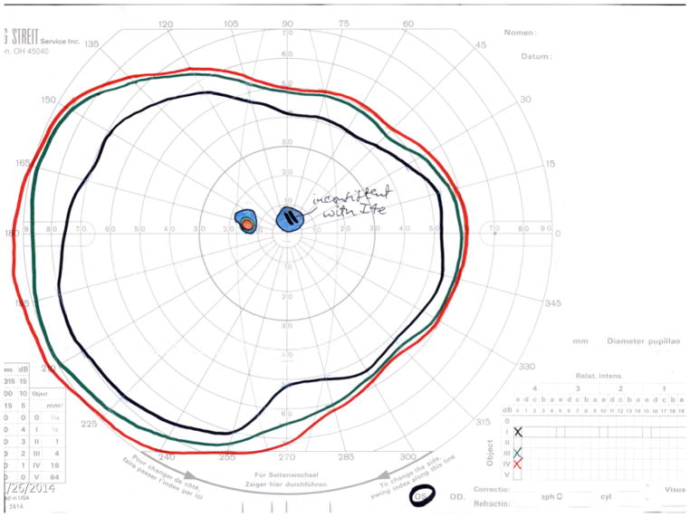
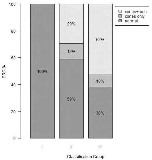
Sixty-five patients with clinical diagnosis and proven pathogenic variants in the ABCA4 gene. Observational Procedures: The UWF-FAF images were obtained using Optos (200 degrees) and classified into 3 types. Functional testing included kinetic widefield perimetry, full-field electroretinogram (ffERG), and visual acuity (VA). All results were evaluated with respect to UWF-FAF classification.
Classification of UWF-FAF; area comprising the I4e, III4e, and IV4e isopters; ffERG patterns; and VA.
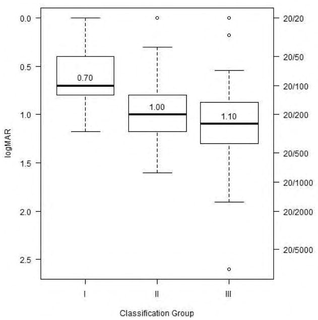
For UWF-FAF, 27 subjects (41.5%) were classified as type I, 17 (26.2%) as type II, and 21 (32.4%) as type III. The area of each isopter correlated inversely with the extent of the disease and all isopters were able to detect differences among UWF-FAF types (IV4e, P = .0013; III4e, P = .0003; I4e, P < .0001 = 3.93e). ffERG patterns and VA were also different among the 3 UWF-FAF types (P < .001 = 6.61e- and P < .001 = 7.3e, respectively).
Patients with widespread disease presented with more constriction of peripheral visual fields and had more dysfunction on ffERG and worse VA compared to patients with disease confined to the macula. UWF-FAF images may provide information for estimating peripheral and central visual function in STGD.
评估ABCA4型斯塔加特病(STGD)患者超广角眼底自发荧光(UWF-FAF)的疾病范围,并将这些数据与功能转归指标相关联。
回顾性横断面研究。
地点:密歇根大学凯洛格眼科中心。
65例临床诊断且ABCA4基因存在已证实的致病变异的患者。观察程序:使用Optos(200度)获取UWF-FAF图像并分为3种类型。功能测试包括动态超广角视野检查、全视野视网膜电图(ffERG)和视力(VA)。所有结果均根据UWF-FAF分类进行评估。
UWF-FAF分类;包含I4e、III4e和IV4e等视线的区域;ffERG模式;以及VA。
对于UWF-FAF,27名受试者(41.5%)被分类为I型,17名(26.2%)为II型,21名(32.4%)为III型。每个等视线的面积与疾病范围呈负相关,并且所有等视线都能够检测出UWF-FAF各类型之间的差异(IV4e,P = 0.0013;III4e,P = 0.0003;I4e,P < 0.0001 = 3.93e)。3种UWF-FAF类型之间的ffERG模式和VA也存在差异(分别为P < 0.001 = 6.61e-和P < 0.001 = 7.3e)。
与疾病局限于黄斑的患者相比,患有广泛疾病的患者周边视野收缩更明显,ffERG功能障碍更严重,VA更差。UWF-FAF图像可能为评估STGD患者的周边和中心视觉功能提供信息。