Department of Medical Joint Materials, Kagoshima University, Kagoshima, Japan.
Department of Orthopaedic Surgery, Kagoshima University, Kagoshima, Japan.
Sci Rep. 2017 Oct 18;7(1):13494. doi: 10.1038/s41598-017-13994-w.
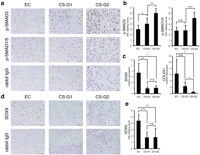
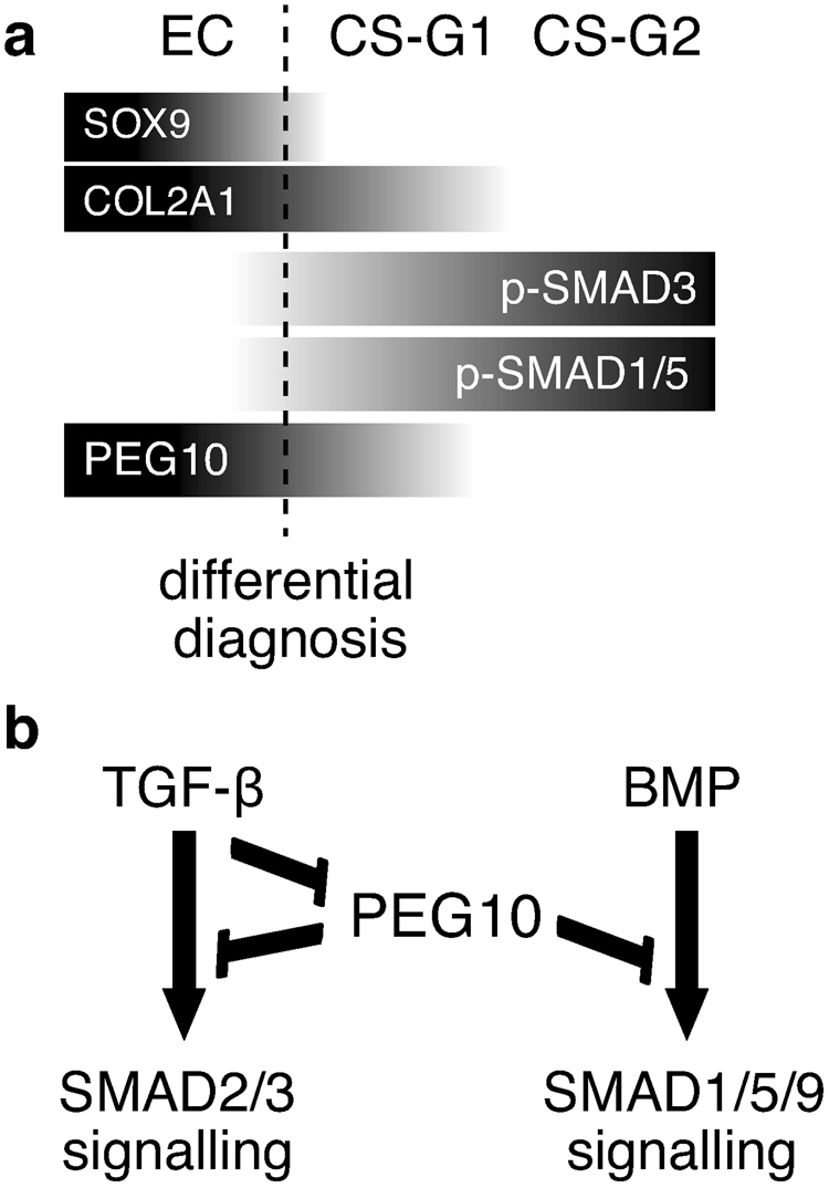
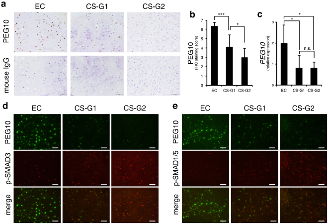
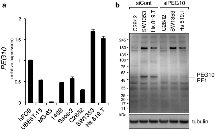
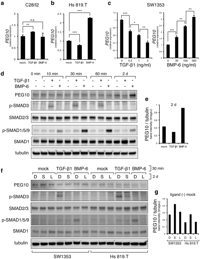
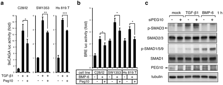
Histological distinction between enchondroma and chondrosarcoma is difficult because of a lack of definitive biomarkers. Here, we found highly active transforming growth factor-β (TGF-β) and bone morphogenetic protein (BMP) signalling in human chondrosarcomas compared with enchondromas by immunohistochemistry of phosphorylated SMAD3 and SMAD1/5. In contrast, the chondrogenic master regulator SOX9 was dramatically down-regulated in grade 1 chondrosarcoma. Paternally expressed gene 10 (PEG10) was identified by microarray analysis as a gene overexpressed in chondrosarcoma SW1353 and Hs 819.T cells compared with C28/I2 normal chondrocytes, while TGF-β1 treatment, mimicking higher grade tumour conditions, suppressed PEG10 expression. Enchondroma samples exhibited stronger expression of PEG10 compared with chondrosarcomas, suggesting a negative association of PEG10 with malignant cartilage tumours. In chondrosarcoma cell lines, application of the TGF-β signalling inhibitor, SB431542, increased the protein level of PEG10. Reporter assays revealed that PEG10 repressed TGF-β and BMP signalling, which are both SMAD pathways, whereas PEG10 knockdown increased the level of phosphorylated SMAD3 and SMAD1/5/9. Our results indicate that mutually exclusive expression of PEG10 and phosphorylated SMADs in combination with differentially expressed SOX9 is an index to distinguish between enchondroma and chondrosarcoma, while PEG10 and TGF-β signalling are mutually inhibitory in chondrosarcoma cells.
软骨母细胞瘤和软骨肉瘤之间的组织学区别因缺乏明确的生物标志物而难以区分。通过对磷酸化 SMAD3 和 SMAD1/5 的免疫组织化学染色,我们发现与软骨母细胞瘤相比,人软骨肉瘤中 TGF-β 和 BMP 信号具有高度活性。相比之下,在 1 级软骨肉瘤中,软骨形成的主调节因子 SOX9 被显著下调。通过微阵列分析鉴定出父系表达基因 10(PEG10)是软骨肉瘤 SW1353 和 Hs 819.T 细胞中过度表达的基因,与 C28/I2 正常软骨细胞相比,而 TGF-β1 处理,模拟更高等级的肿瘤条件,抑制 PEG10 表达。软骨母细胞瘤样本中 PEG10 的表达强于软骨肉瘤,提示 PEG10 与恶性软骨肿瘤呈负相关。在软骨肉瘤细胞系中,应用 TGF-β 信号抑制剂 SB431542 可增加 PEG10 的蛋白水平。报告基因检测显示,PEG10 抑制 TGF-β 和 BMP 信号,这两种信号均为 SMAD 通路,而 PEG10 敲低增加了磷酸化 SMAD3 和 SMAD1/5/9 的水平。我们的研究结果表明,PEG10 和磷酸化 SMAD 表达的互斥表达以及 SOX9 的差异表达是区分软骨母细胞瘤和软骨肉瘤的指标,而 PEG10 和 TGF-β 信号在软骨肉瘤细胞中是相互抑制的。