Program in Microbiology, Institute of Medical Sciences, University of Aberdeen, Aberdeen, United Kingdom.
School of Molecular Biosciences, College of Veterinary Medicine, Washington State University, Pullman, WA, United States.
Front Cell Infect Microbiol. 2017 Oct 10;7:438. doi: 10.3389/fcimb.2017.00438. eCollection 2017.
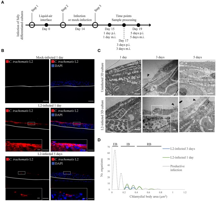
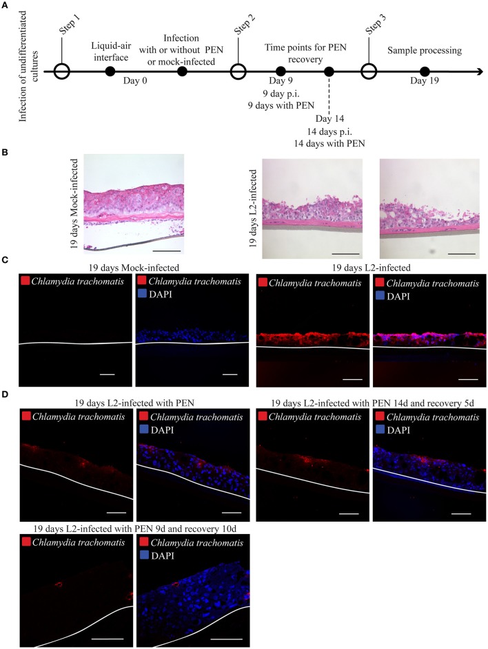
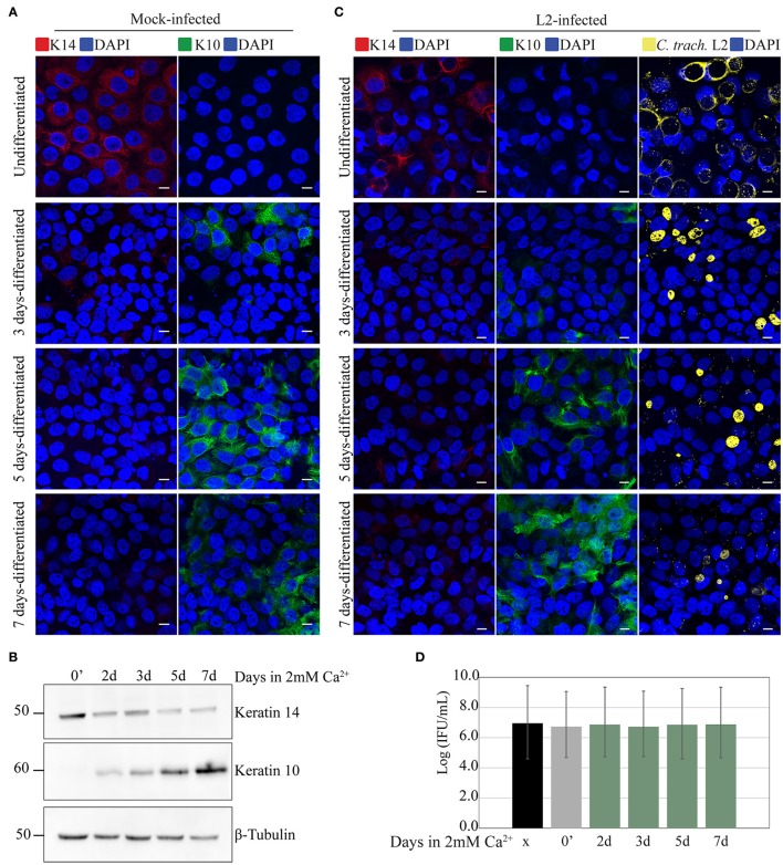
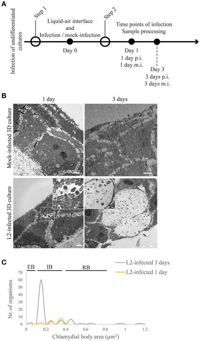
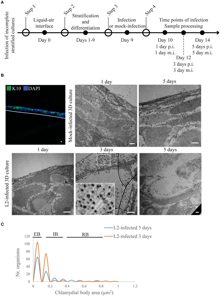
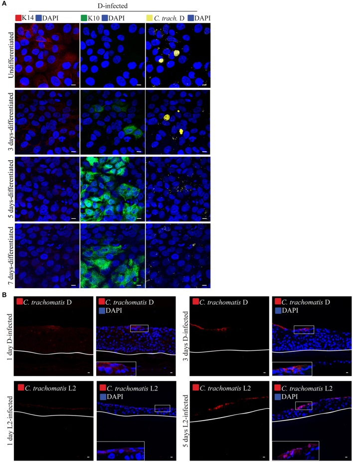
infection targets the mucosal epithelium, where squamous and columnar epithelia can be found. Research on -epithelia interaction has predominantly focused on columnar epithelia, with very little known on how interacts with the squamous epithelium. The stratification and differentiation processes found in the squamous epithelium might influence chlamydial growth and infection dissemination. For this reason, three-dimensional (3D) organotypic stratified squamous epithelial cultures were adapted to mimic the stratified squamous epithelium and chlamydial infection was characterized. infection in monolayers and 3D cultures were monitored by immunofluorescence and transmission electron microscopy to evaluate inclusion growth and chlamydial interconversion between elementary and reticulate body. We observed that the stratified epithelium varied in susceptibility to serovars L2 and D infection. The undifferentiated basal cells were susceptible to infection by both serovars, while the terminally differentiated upper layers were resistant. The differentiating suprabasal cells exhibited different susceptibilities to serovars L2 and D, with the latter unable to establish a successful infection in this layer. Mature elementary body-containing inclusions were much more prevalent in these permissive basal layers, while the uppermost differentiated layers consistently harbored very few reticulate bodies with no elementary bodies, indicative of severely limited bacterial replication and development. For serovar D, the differentiation state of the host cell was a determining factor, as calcium-induced differentiation of cells in a monolayer negatively affected growth of this serovar, in contrast to serovar L2. The apparent completion of the developmental cycle in the basal layers of the 3D cultures correlated with the greater degree of dissemination within and the level of disruption of the stratified epithelium. Our studies indicate that the squamous epithelium is a suboptimal environment for growth, and thus potentially contributing to the protection of the lower genital tract from infection. The relatively more fastidious serovar D exhibited more limited growth than the faster-growing and more invasive L2 strain. However, if given access to the more hospitable basal cell layer, both strains were able to produce mature inclusions, replicate, and complete their developmental cycle.
感染靶标是黏膜上皮,其中可以发现鳞状上皮和柱状上皮。对 -上皮相互作用的研究主要集中在柱状上皮上,而对于如何与鳞状上皮相互作用知之甚少。在鳞状上皮中发现的分层和分化过程可能会影响衣原体的生长和感染传播。出于这个原因,适应了三维(3D)器官样分层鳞状上皮培养以模拟分层鳞状上皮,并对衣原体感染进行了特征描述。通过免疫荧光和透射电子显微镜监测单层和 3D 培养物中的感染,以评估包涵体的生长和衣原体在原体和网状体之间的相互转化。我们观察到,分层上皮对 L2 和 D 血清型的感染易感性不同。未分化的基底细胞对两种血清型均易感,而终末分化的上层则具有抗性。分化的基底上层细胞对 L2 和 D 血清型的易感性不同,后者在该层中无法建立成功的感染。在这些允许的基底层中,成熟的包含原体的包涵体更为普遍,而上层分化的层始终带有很少的网状体而没有原体,表明细菌复制和发育受到严重限制。对于 D 血清型,宿主细胞的分化状态是一个决定因素,因为单层细胞中的钙诱导分化对该血清型的生长产生负面影响,而与 L2 血清型相反。3D 培养物中基底层中明显完成发育周期与在分层上皮内的更大程度的扩散和破坏水平相关。我们的研究表明,鳞状上皮是生长的次优环境,因此可能有助于保护下生殖道免受感染。相对更挑剔的 D 血清型比生长更快、侵袭性更强的 L2 株生长受到更有限的限制。然而,如果能够进入更适合的基底细胞层,两种菌株都能够产生成熟的包涵体、复制并完成其发育周期。