Manning Kathryn Y, Schranz Amy, Bartha Robert, Dekaban Gregory A, Barreira Christy, Brown Arthur, Fischer Lisa, Asem Kevin, Doherty Timothy J, Fraser Douglas D, Holmes Jeff, Menon Ravi S
From the Department of Medical Biophysics (K.Y.M., A.S., R.B., R.S.M.), Department of Microbiology and Immunology (G.A.D., C.B.), Department of Anatomy and Cell Biology (A.B), Department of Physical Medicine and Rehabilitation (T.J.D.), and School of Occupational Therapy (J.H.), University of Western Ontario; Centre for Functional and Metabolic Mapping (K.Y.M., R.B., R.S.M.) and Molecular Medicine (G.A.D., C.B., A.B.), Robarts Research Institute; Primary Care Sport Medicine (L.F., K.A.), Family Medicine, Fowler Kennedy Sport Medicine; and Paediatrics Critical Care Medicine (D.D.F.), London Health Sciences Centre, London, Ontario, Canada.
Neurology. 2017 Nov 21;89(21):2157-2166. doi: 10.1212/WNL.0000000000004669. Epub 2017 Oct 25.
To determine whether multiparametric MRI data can provide insight into the acute and long-lasting neuronal sequelae after a concussion in adolescent athletes.
Players were recruited from Bantam hockey leagues in which body checking is first introduced (male, age 11-14 years). Clinical measures, diffusion metrics, resting-state network and region-to-region functional connectivity patterns, and magnetic resonance spectroscopy absolute metabolite concentrations were analyzed from an independent, age-matched control group of hockey players (n = 26) and longitudinally in concussed athletes within 24 to 72 hours (n = 17) and 3 months (n = 14) after a diagnosed concussion.
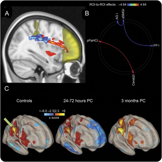
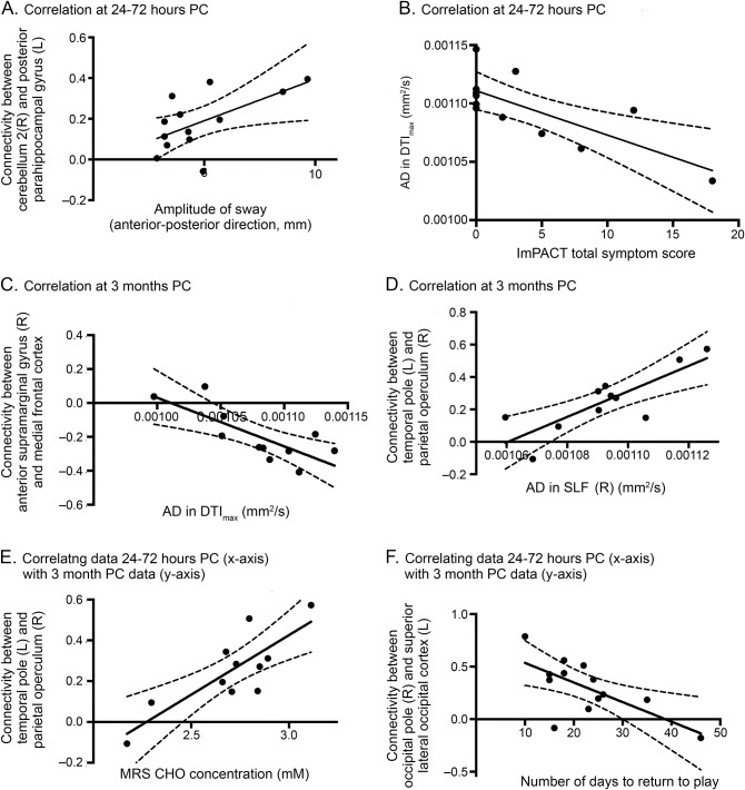
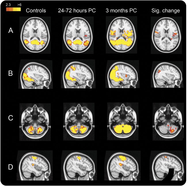
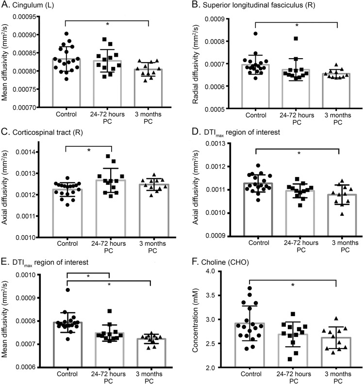
There were diffusion abnormalities within multiple white matter tracts, functional hyperconnectivity, and decreases in choline 3 months after concussion. Tract-specific spatial statistics revealed a large region along the superior longitudinal fasciculus with the largest decreases in diffusivity measures, which significantly correlated with clinical deficits. This region also spatially intersected with probabilistic tracts connecting cortical regions where we found acute functional connectivity changes. Hyperconnectivity patterns at 3 months after concussion were present only in players with relatively less severe clinical outcomes, higher choline concentrations, and diffusivity indicative of relatively less axonal disruption.
Changes persisted well after players' clinical scores had returned to normal and they had been cleared to return to play. Ongoing white matter maturation may make adolescent athletes particularly vulnerable to brain injury, and they may require extended recovery periods. The consequences of early brain injury for ongoing brain development and risk of more serious conditions such as second impact syndrome or neural degenerative processes need to be elucidated.
确定多参数MRI数据能否为青少年运动员脑震荡后的急性和长期神经元后遗症提供深入见解。
从首次引入身体冲撞的少年曲棍球联盟招募运动员(男性,11 - 14岁)。对一组独立的、年龄匹配的曲棍球运动员对照组(n = 26)进行临床测量、扩散指标、静息态网络和区域间功能连接模式以及磁共振波谱绝对代谢物浓度分析,并对确诊脑震荡后24至72小时(n = 17)和3个月(n = 14)的脑震荡运动员进行纵向分析。
脑震荡后3个月,多个白质束内存在扩散异常、功能连接增强以及胆碱减少。特定束的空间统计显示,沿上纵束有一大区域扩散率测量值下降最大,这与临床缺陷显著相关。该区域在空间上还与连接皮质区域的概率束相交,在这些皮质区域我们发现了急性功能连接变化。脑震荡后3个月的功能连接增强模式仅出现在临床结果相对较轻、胆碱浓度较高且扩散率表明轴突损伤相对较轻的运动员中。
在运动员临床评分恢复正常并被允许重返比赛后,这些变化仍持续存在。持续的白质成熟可能使青少年运动员特别容易受到脑损伤,他们可能需要更长的恢复期。早期脑损伤对持续脑发育的影响以及诸如二次撞击综合征或神经退行性疾病等更严重病症的风险需要进一步阐明。