Blair Norman P, Wanek Justin, Felder Anthony E, Joslin Charlotte E, Kresovich Jacob K, Lim Jennifer I, Chau Felix Y, Leiderman Yannek, Shahidi Mahnaz
Department of Ophthalmology and Visual Sciences, University of Illinois at Chicago, Chicago, Illinois, United States.
Division of Epidemiology and Biostatistics, School of Public Health, University of Illinois at Chicago, Chicago, Illinois, United States.
Invest Ophthalmol Vis Sci. 2017 Oct 1;58(12):5556-5563. doi: 10.1167/iovs.17-21934.
To test the hypothesis that retinal vascular diameter and hemoglobin oxygen saturation alterations, according to stages of diabetic retinopathy (DR), are discernible with a commercially available scanning laser ophthalmoscope (SLO).
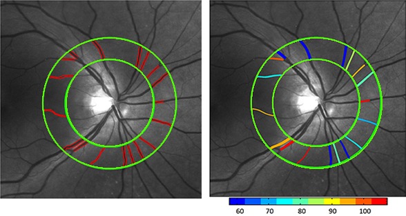
One hundred eighty-one subjects with no diabetes (No DM), diabetes with no DR (No DR), nonproliferative DR (NPDR), or proliferative DR (PDR, all had photocoagulation) underwent imaging with an SLO with dual lasers (532 nm and 633 nm). Customized image analysis software determined the diameters of retinal arteries and veins (DA and DV) and central retinal artery and vein equivalents (CRAE and CRVE). Oxygen saturations of hemoglobin in arteries and veins (SO2A and SO2V) were estimated from optical densities of vessels on images at the two wavelengths. Statistical models were generated by adjusting for effects of sex, race, age, eye, and fundus pigmentation.
DA, CRAE, and CRVE were reduced in PDR compared to No DM (P ≤ 0.03). DV and CRVE were similar between No DM and No DR, but they were higher in NPDR than No DR (P ≤ 0.01). Effect of stage of disease on SO2A differed by race, being increased relative to No DM in NPDR and PDR in Hispanic participants only (P ≤ 0.02). Relative to No DM, SO2V was increased in NPDR and PDR (P ≤ 0.05).
Alterations in retinal vascular diameters and SO2 by diabetic retinopathy stage can be detected with a widely available SLO, and covariates such as race can influence the results.
验证以下假设,即使用商用扫描激光检眼镜(SLO)可辨别出糖尿病视网膜病变(DR)各阶段的视网膜血管直径和血红蛋白氧饱和度变化。
181名受试者,包括无糖尿病者(无DM)、患有糖尿病但无DR者(无DR)、非增殖性DR(NPDR)患者或增殖性DR(PDR,均接受了光凝治疗),使用双激光(532纳米和633纳米)的SLO进行成像。定制的图像分析软件确定视网膜动脉和静脉的直径(DA和DV)以及视网膜中央动脉和静脉等效直径(CRAE和CRVE)。根据两个波长图像上血管的光密度估算动脉和静脉中血红蛋白的氧饱和度(SO2A和SO2V)。通过调整性别、种族、年龄、眼睛和眼底色素沉着的影响来生成统计模型。
与无DM相比,PDR患者的DA、CRAE和CRVE减小(P≤0.03)。无DM和无DR患者的DV和CRVE相似,但NPDR患者的DV和CRVE高于无DR患者(P≤0.01)。疾病阶段对SO2A的影响因种族而异,仅在西班牙裔参与者的NPDR和PDR中相对于无DM增加(P≤0.02)。相对于无DM,NPDR和PDR患者的SO2V增加(P≤0.05)。
使用广泛可用的SLO可以检测出糖尿病视网膜病变各阶段视网膜血管直径和SO2的变化,种族等协变量会影响结果。