Department of Biochemistry and Microbiology, Oklahoma State University-Center for Health Sciences, Tulsa, OK 74107, United States.
Immatics US Inc, Houston, TX 77077, United States.
World J Gastroenterol. 2017 Oct 7;23(37):6802-6816. doi: 10.3748/wjg.v23.i37.6802.
To investigate gender-specific liver estrogen receptor (ER) expression in normal subjects and patients with hepatitis C virus (HCV)-related cirrhosis and hepatocellular carcinoma (HCC).
Liver tissues from normal donors and patients diagnosed with HCV-related cirrhosis and HCV-related HCC were obtained from the NIH Liver Tissue and Cell Distribution System. The expression of ER subtypes, ERα and ERβ, were evaluated by Western blotting and real-time RT-PCR. The subcellular distribution of ERα and ERβ was further determined in nuclear and cytoplasmic tissue lysates along with the expression of inflammatory [activated NF-κB and IκB-kinase (IKK)] and oncogenic (cyclin D1) markers by Western blotting and immunohistochemistry. The expression of ERα and ERβ was correlated with the expression of activated NF-κB, activated IKK and cyclin D1 by Spearman's correlation.
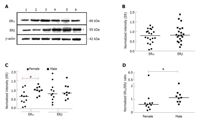
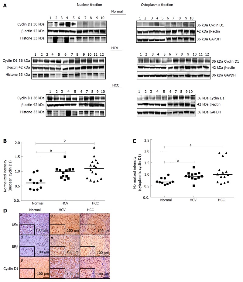
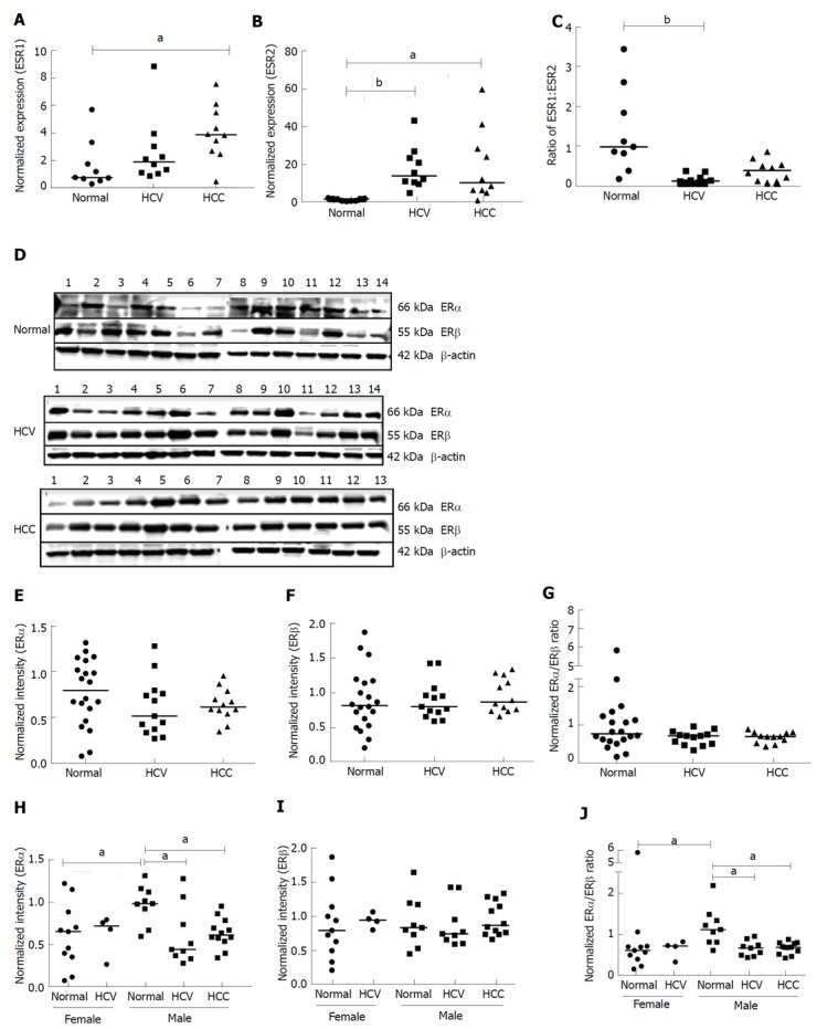
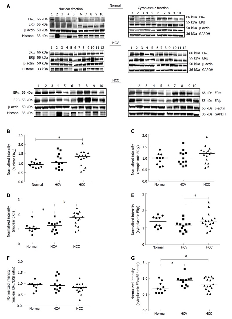
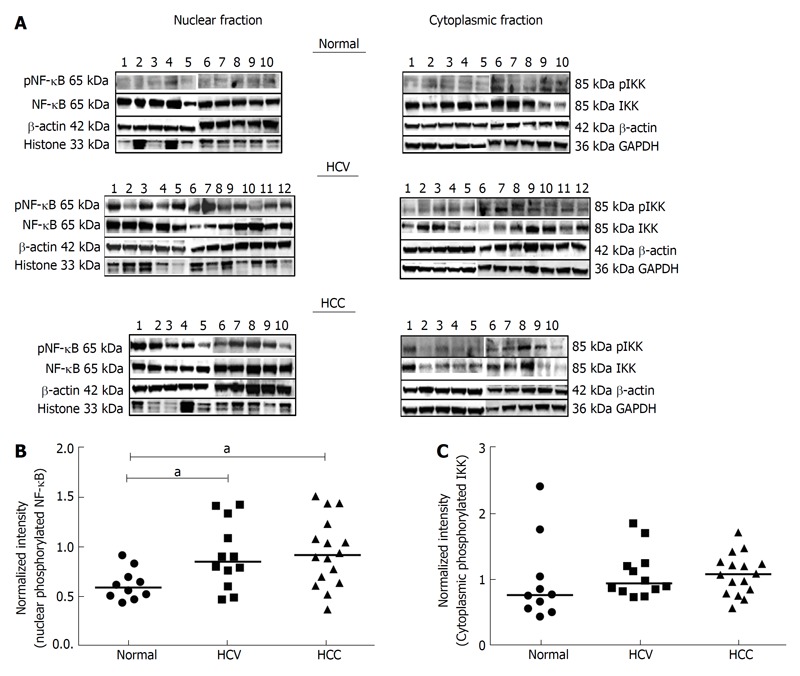
Both ER subtypes were expressed in normal livers but male livers showed significantly higher expression of ERα than females ( < 0.05). We observed significantly higher mRNA expression of ERα in HCV-related HCC liver tissues as compared to normals ( < 0.05) and ERβ in livers of HCV-related cirrhosis and HCV-related HCC subjects ( < 0.05). At the protein level, there was a significantly higher expression of nuclear ERα in livers of HCV-related HCC patients and nuclear ERβ in HCV-related cirrhosis patients as compared to normals ( < 0.05). Furthermore, we observed a significantly higher expression of phosphorylated NF-κB and cyclin D1 in diseased livers ( < 0.05). There was a positive correlation between the expression of nuclear ER subtypes and nuclear cyclin D1 and a negative correlation between cytoplasmic ER subtypes and cytoplasmic phosphorylated IKK in HCV-related HCC livers. These findings suggest that dysregulated expression of ER subtypes following chronic HCV-infection may contribute to the progression of HCV-related cirrhosis to HCV-related HCC.
Gender differences were observed in ERα expression in normal livers. Alterations in ER subtype expression observed in diseased livers may influence gender-related disparity in HCV-related pathogenesis.
研究正常人和丙型肝炎病毒(HCV)相关肝硬化和肝细胞癌(HCC)患者肝雌激素受体(ER)的性别特异性表达。
从 NIH 肝脏组织和细胞分配系统获得正常供体和诊断为 HCV 相关肝硬化和 HCV 相关 HCC 的患者的肝组织。通过 Western 印迹和实时 RT-PCR 评估 ER 亚型 ERα和 ERβ的表达。通过 Western 印迹和免疫组织化学进一步确定 ERα和 ERβ在核和细胞质组织裂解物中的亚细胞分布,以及炎症(激活的 NF-κB 和 IκB 激酶(IKK))和致癌(细胞周期蛋白 D1)标志物的表达。通过 Spearman 相关性分析将 ERα和 ERβ的表达与激活的 NF-κB、激活的 IKK 和细胞周期蛋白 D1 的表达相关联。
两种 ER 亚型在正常肝脏中均有表达,但男性肝脏中 ERα的表达明显高于女性(<0.05)。与正常肝脏相比,我们观察到 HCV 相关 HCC 肝脏组织中 ERα 的 mRNA 表达明显升高(<0.05),而 HCV 相关肝硬化和 HCC 患者的 ERβ表达升高(<0.05)。在蛋白质水平上,与正常肝脏相比,HCV 相关 HCC 患者的核 ERα和 HCV 相关肝硬化患者的核 ERβ表达明显升高(<0.05)。此外,我们观察到病变肝脏中磷酸化 NF-κB 和细胞周期蛋白 D1 的表达明显升高(<0.05)。在 HCV 相关 HCC 肝脏中,核 ER 亚型的表达与核细胞周期蛋白 D1呈正相关,细胞质 ER 亚型的表达与细胞质磷酸化 IKK 呈负相关。这些发现表明,慢性 HCV 感染后 ER 亚型表达的失调可能导致 HCV 相关肝硬化向 HCV 相关 HCC 的进展。
在正常肝脏中观察到 ERα表达存在性别差异。在病变肝脏中观察到的 ER 亚型表达的改变可能会影响 HCV 相关发病机制中的性别差异。