Ippolito Davide, Casiraghi Alessandra Silvia, Franzesi Cammillo Talei, Fior Davide, Meloni Franca, Sironi Sandro
Department of Diagnostic Radiology, "San Gerardo" Hospital, 20900 Monza, Italy.
School of Medicine, University of Milano-Bicocca, 20900 Monza, Italy.
World J Gastrointest Oncol. 2017 Oct 15;9(10):423-430. doi: 10.4251/wjgo.v9.i10.423.
To compare radiation dose and image quality of low-dose computed tomography (CT) protocol combined with hybrid-iterative reconstruction algorithm with standard-dose CT examinations for follow-up of oncologic patients.
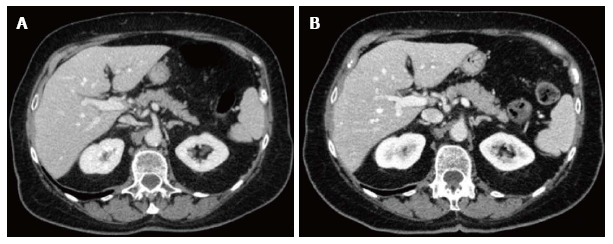
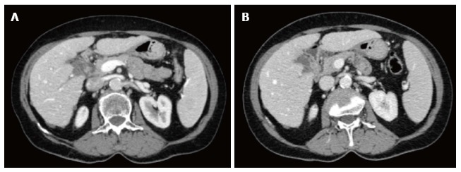
Fifty-one patients with known malignant diseases which underwent, during clinical follow-up, both standard-dose and low-dose whole-body CT scans were enrolled. Low-dose CT was performed on 256-row scanner, with 120 kV and automated mA modulation, and iterative reconstruction algorithm. Standard-dose CT was performed on 16-rows scanner, with 120 kV, 200-400 mAs (depending on patient weight). We evaluated density values and signal-to-noise ratio, along with image noise (SD), sharpness and diagnostic quality with 4-point scale.
Density values in liver, spleen and aorta were higher in low-dose images (liver 112.55 HU 103.90 HU, < 0.001), as SD values in liver and spleen (liver 16.81 14.41). Volumetric-Computed-Tomographic-Dose-Index (CTDIvol) and Dose-Length-Product (DLP) were significantly lower in low-dose CT as compared to standard-dose (DLP 1025.6 mGycm 1429.2 mGycm, < 0.001) with overall dose reduction of 28.9%. Qualitative analysis did not reveal significant differences in image noise and diagnostic quality.
Automatic tube-current modulation combined with hybrid-iterative algorithm allows radiation dose reduction of 28.9% without loss of diagnostic quality, being useful in reducing dose exposure in oncologic patients.
比较低剂量计算机断层扫描(CT)方案联合混合迭代重建算法与标准剂量CT检查在肿瘤患者随访中的辐射剂量和图像质量。
纳入51例已知恶性疾病患者,这些患者在临床随访期间接受了标准剂量和低剂量全身CT扫描。低剂量CT在256排扫描仪上进行,管电压120 kV,自动毫安调制,并采用迭代重建算法。标准剂量CT在16排扫描仪上进行,管电压120 kV,管电流200 - 400 mAs(根据患者体重而定)。我们评估了密度值、信噪比,以及图像噪声(标准差)、清晰度和诊断质量,并采用4分制进行评分。
低剂量图像中肝脏、脾脏和主动脉的密度值较高(肝脏112.55 HU对103.90 HU,<0.001),肝脏和脾脏的标准差也较高(肝脏16.81对14.41)。与标准剂量相比,低剂量CT的容积计算机断层扫描剂量指数(CTDIvol)和剂量长度乘积(DLP)显著降低(DLP 1025.6 mGy·cm对1429.2 mGy·cm,<0.001),总体剂量降低了28.9%。定性分析未发现图像噪声和诊断质量有显著差异。
自动管电流调制联合混合迭代算法可使辐射剂量降低28.9%,且不损失诊断质量,有助于减少肿瘤患者的剂量暴露。