• 文献检索
  • 文档翻译
  • 深度研究
  • 学术资讯
  • Suppr Zotero 插件Zotero 插件
  • 邀请有礼
  • 套餐&价格
  • 历史记录
应用&插件
Suppr Zotero 插件Zotero 插件浏览器插件Mac 客户端Windows 客户端微信小程序
定价
高级版会员购买积分包购买API积分包
服务
文献检索文档翻译深度研究API 文档MCP 服务
关于我们
关于 Suppr公司介绍联系我们用户协议隐私条款
关注我们

Suppr 超能文献

核心技术专利:CN118964589B侵权必究
粤ICP备2023148730 号-1Suppr @ 2026

文献检索

告别复杂PubMed语法,用中文像聊天一样搜索,搜遍4000万医学文献。AI智能推荐,让科研检索更轻松。

立即免费搜索

文件翻译

保留排版,准确专业,支持PDF/Word/PPT等文件格式,支持 12+语言互译。

免费翻译文档

深度研究

AI帮你快速写综述,25分钟生成高质量综述,智能提取关键信息,辅助科研写作。

立即免费体验

侵袭性卵巢肿瘤产生的恶性腹水的促血管生成能力。

The Proangiogenic Capabilities of Malignant Ascites Generated by Aggressive Ovarian Tumors.

机构信息

Department of Hypertensiology, Angiology and Internal Medicine, Poznań University of Medical Sciences, Długa 1/2 Str., 61-848 Poznań, Poland.

Division of Gynecological Surgery, Poznań University of Medical Sciences, Polna 33 Str., 60-535 Poznań, Poland.

出版信息

Biomed Res Int. 2017;2017:2592496. doi: 10.1155/2017/2592496. Epub 2017 Sep 20.

DOI:10.1155/2017/2592496
PMID:29085834
原文链接:https://pmc.ncbi.nlm.nih.gov/articles/PMC5632494/
Abstract

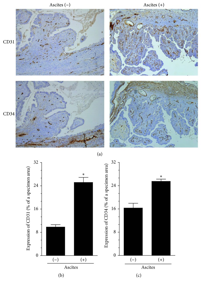
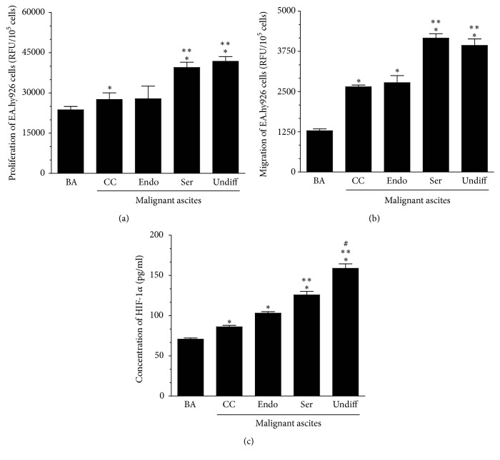
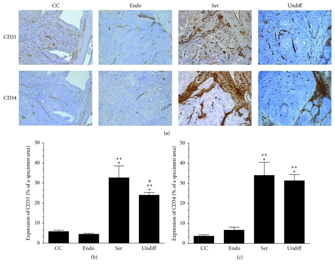
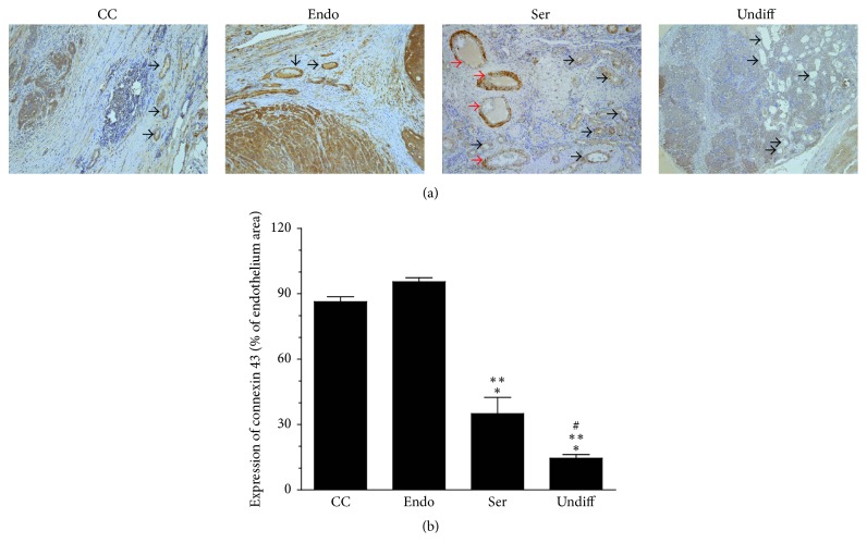
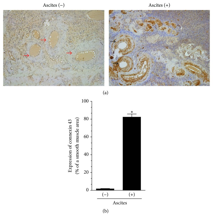
Here we examined whether malignant ascites may determine ovarian tumor angiogenesis, and if so whether ascites generated by highly aggressive serous and undifferentiated cancers are more proangiogenic than those from less aggressive clear cell and endometrioid tumors. Angiogenesis was analyzed according to expression of CD31, CD34, and connexin 43. Proliferation and migration of endothelial cells were tested using fluorescence-based methods. The quantification of angiogenic agents and hypoxia-inducible factor 1 (HIF-1) was performed using specific immunoassays. Results showed that the expression of CD31 and CD34 in serous and undifferentiated tumors was greater, whereas endothelial expression of connexin 43 was lower than in clear cell and endometrioid lesions. Serous cancers that formed in the presence of ascites displayed increased expression of connexin 43 in vascular smooth muscles as compared with tumors developed in the fluid's absence. Endothelial cells exposed to ascites from serous and undifferentiated tumors proliferated and migrated more vigorously than cells subjected to ascites from clear cell and endometrioid cancers. They also exhibited an increased level of HIF-1 and produced increased amounts of multiple proangiogenic agents. Our results indicate that high vascularization of aggressive ovarian tumors may be associated with profound angiogenic capabilities of ascites generated by these tumors.

摘要

在这里,我们研究了恶性腹水是否会决定卵巢肿瘤的血管生成,如果是,那么高度侵袭性的浆液性和未分化癌症产生的腹水是否比低度侵袭性的透明细胞和子宫内膜样肿瘤产生的腹水更具血管生成能力。血管生成根据 CD31、CD34 和连接蛋白 43 的表达进行分析。使用荧光法测试内皮细胞的增殖和迁移。使用特定的免疫测定法对血管生成剂和缺氧诱导因子 1(HIF-1)的定量进行了检测。结果表明,浆液性和未分化肿瘤中 CD31 和 CD34 的表达更高,而内皮细胞连接蛋白 43 的表达低于透明细胞和子宫内膜样病变。在存在腹水的情况下形成的浆液性癌症与在没有腹水的情况下形成的肿瘤相比,血管平滑肌中连接蛋白 43 的表达增加。与暴露于透明细胞和子宫内膜样癌腹水的细胞相比,暴露于浆液性和未分化肿瘤腹水的内皮细胞增殖和迁移更为活跃。它们还表现出更高水平的 HIF-1,并产生更多的多种促血管生成剂。我们的结果表明,侵袭性卵巢肿瘤的高血管化可能与这些肿瘤产生的腹水具有深远的血管生成能力有关。

https://cdn.ncbi.nlm.nih.gov/pmc/blobs/a4c9/5632494/7e2f1b5bc853/BMRI2017-2592496.005.jpg
https://cdn.ncbi.nlm.nih.gov/pmc/blobs/a4c9/5632494/f93c87752de2/BMRI2017-2592496.001.jpg
https://cdn.ncbi.nlm.nih.gov/pmc/blobs/a4c9/5632494/63b15191b505/BMRI2017-2592496.002.jpg
https://cdn.ncbi.nlm.nih.gov/pmc/blobs/a4c9/5632494/68aa7710ed0f/BMRI2017-2592496.003.jpg
https://cdn.ncbi.nlm.nih.gov/pmc/blobs/a4c9/5632494/ec309cec8d67/BMRI2017-2592496.004.jpg
https://cdn.ncbi.nlm.nih.gov/pmc/blobs/a4c9/5632494/7e2f1b5bc853/BMRI2017-2592496.005.jpg

相似文献

1
The Proangiogenic Capabilities of Malignant Ascites Generated by Aggressive Ovarian Tumors.侵袭性卵巢肿瘤产生的恶性腹水的促血管生成能力。
Biomed Res Int. 2017;2017:2592496. doi: 10.1155/2017/2592496. Epub 2017 Sep 20.
2
Malignant ascites determine the transmesothelial invasion of ovarian cancer cells.恶性腹水决定了卵巢癌细胞的跨间皮侵袭。
Int J Biochem Cell Biol. 2017 Nov;92:6-13. doi: 10.1016/j.biocel.2017.09.002. Epub 2017 Sep 6.
3
Biochemical composition of malignant ascites determines high aggressiveness of undifferentiated ovarian tumors.恶性腹水的生化组成决定了未分化卵巢肿瘤的高侵袭性。
Med Oncol. 2016 Aug;33(8):94. doi: 10.1007/s12032-016-0810-4. Epub 2016 Jul 18.
4
Thrombospondin-1 and -2 messenger RNA expression in epithelial ovarian tumor.血小板反应蛋白-1和-2信使核糖核酸在上皮性卵巢肿瘤中的表达
Anticancer Res. 2001 Jul-Aug;21(4B):2983-7.
5
CXCL12 and vascular endothelial growth factor synergistically induce neoangiogenesis in human ovarian cancers.CXCL12与血管内皮生长因子协同诱导人卵巢癌新生血管形成。
Cancer Res. 2005 Jan 15;65(2):465-72.
6
Soluble FLT-1 expression suppresses carcinomatous ascites in nude mice bearing ovarian cancer.可溶性FLT-1表达可抑制荷卵巢癌裸鼠的癌性腹水。
Cancer Res. 2002 Apr 1;62(7):2019-23.
7
Vascular endothelial growth factor activating matrix metalloproteinase in ascitic fluid during peritoneal dissemination of ovarian cancer.血管内皮生长因子在卵巢癌腹膜播散过程中激活腹水中的基质金属蛋白酶。
Oncol Rep. 2003 Jan-Feb;10(1):89-95.
8
Expression of hypoxia-inducible factor 1alpha, hypoxia-inducible factor 2alpha, and von Hippel-Lindau protein in epithelial ovarian neoplasms and allelic loss of von Hippel-Lindau gene: nuclear expression of hypoxia-inducible factor 1alpha is an independent prognostic factor in ovarian carcinoma.缺氧诱导因子1α、缺氧诱导因子2α及冯·希佩尔-林道蛋白在上皮性卵巢肿瘤中的表达及冯·希佩尔-林道基因的等位基因缺失:缺氧诱导因子1α的核表达是卵巢癌的独立预后因素。
Hum Pathol. 2007 Sep;38(9):1310-20. doi: 10.1016/j.humpath.2007.02.010. Epub 2007 Jun 6.
9
Vasohibin-2 expressed in human serous ovarian adenocarcinoma accelerates tumor growth by promoting angiogenesis.人浆液性卵巢腺癌中表达的血管抑素-2通过促进血管生成加速肿瘤生长。
Mol Cancer Res. 2012 Sep;10(9):1135-46. doi: 10.1158/1541-7786.MCR-12-0098-T. Epub 2012 Jul 23.
10
Expression of thymidine phosphorylase and vascular endothelial growth factor in epithelial ovarian cancer: correlation with angiogenesis and progression of the tumor.胸苷磷酸化酶和血管内皮生长因子在上皮性卵巢癌中的表达:与肿瘤血管生成及进展的相关性
Anticancer Res. 2000 Sep-Oct;20(5C):3941-9.

引用本文的文献

1
The Multifaceted Role of Connexins in Tumor Microenvironment Initiation and Maintenance.连接蛋白在肿瘤微环境起始与维持中的多方面作用
Biology (Basel). 2023 Jan 28;12(2):204. doi: 10.3390/biology12020204.
2
Clinicopathologic factors associated with short-term survival in advanced epithelial ovarian cancer.与晚期上皮性卵巢癌短期生存相关的临床病理因素
Transl Cancer Res. 2019 Oct;8(6):2396-2404. doi: 10.21037/tcr.2019.09.53.
3
Malignant Ascites Promote Adhesion of Ovarian Cancer Cells to Peritoneal Mesothelium and Fibroblasts.

本文引用的文献

1
Biochemical composition of malignant ascites determines high aggressiveness of undifferentiated ovarian tumors.恶性腹水的生化组成决定了未分化卵巢肿瘤的高侵袭性。
Med Oncol. 2016 Aug;33(8):94. doi: 10.1007/s12032-016-0810-4. Epub 2016 Jul 18.
2
Tumour Angiogenesis and Angiogenic Inhibitors: A Review.肿瘤血管生成与血管生成抑制剂:综述
J Clin Diagn Res. 2015 Jun;9(6):XE01-XE05. doi: 10.7860/JCDR/2015/12016.6135. Epub 2015 Jun 1.
3
Ovarian cancer ascites enhance the migration of patient-derived peritoneal mesothelial cells via cMet pathway through HGF-dependent and -independent mechanisms.
恶性腹水促进卵巢癌细胞与腹膜间皮细胞和成纤维细胞的黏附。
Int J Mol Sci. 2021 Apr 19;22(8):4222. doi: 10.3390/ijms22084222.
4
The Epithelial-Mesenchymal Transition Initiated by Malignant Ascites Underlies the Transmesothelial Invasion of Ovarian Cancer Cells.恶性腹水引发的上皮-间充质转化是卵巢癌细胞穿破间皮的基础。
Int J Mol Sci. 2019 Jan 2;20(1):137. doi: 10.3390/ijms20010137.
卵巢癌腹水通过 HGF 依赖性和非依赖性机制通过 cMet 途径增强患者来源的腹膜间皮细胞的迁移。
Int J Cancer. 2015 Jul 15;137(2):289-98. doi: 10.1002/ijc.29385. Epub 2014 Dec 18.
4
Bevacizumab in combination with chemotherapy for the treatment of advanced ovarian cancer: a systematic review.贝伐单抗联合化疗治疗晚期卵巢癌:一项系统评价
J Ovarian Res. 2014 May 19;7:57. doi: 10.1186/1757-2215-7-57. eCollection 2014.
5
The angiogenic activity of ascites in the course of ovarian cancer as a marker of disease progression.腹水的血管生成活性作为卵巢癌疾病进展的标志物。
Dis Markers. 2014;2014:683757. doi: 10.1155/2014/683757. Epub 2014 Jan 23.
6
Recent molecular discoveries in angiogenesis and antiangiogenic therapies in cancer.癌症中血管生成和抗血管生成治疗的最新分子发现。
J Clin Invest. 2013 Aug;123(8):3190-200. doi: 10.1172/JCI70212. Epub 2013 Aug 1.
7
Angiogenesis-related pathways in the pathogenesis of ovarian cancer.卵巢癌发病机制中的血管生成相关途径。
Int J Mol Sci. 2013 Jul 30;14(8):15885-909. doi: 10.3390/ijms140815885.
8
Molecular mechanisms of tumor angiogenesis.肿瘤血管生成的分子机制
Genes Cancer. 2011 Dec;2(12):1085-96. doi: 10.1177/1947601911432334.
9
Significance of vascular endothelial growth factor in growth and peritoneal dissemination of ovarian cancer.血管内皮生长因子在卵巢癌生长和腹膜转移中的意义。
Cancer Metastasis Rev. 2012 Jun;31(1-2):143-62. doi: 10.1007/s10555-011-9337-5.
10
Endothelial expression of autocrine VEGF upon the uptake of tumor-derived microvesicles containing oncogenic EGFR.在摄取含有致癌性表皮生长因子受体(EGFR)的肿瘤衍生微囊泡后,内皮细胞自分泌血管内皮生长因子(VEGF)的表达。
Proc Natl Acad Sci U S A. 2009 Mar 10;106(10):3794-9. doi: 10.1073/pnas.0804543106. Epub 2009 Feb 20.