• 文献检索
  • 文档翻译
  • 深度研究
  • 学术资讯
  • Suppr Zotero 插件Zotero 插件
  • 邀请有礼
  • 套餐&价格
  • 历史记录
应用&插件
Suppr Zotero 插件Zotero 插件浏览器插件Mac 客户端Windows 客户端微信小程序
定价
高级版会员购买积分包购买API积分包
服务
文献检索文档翻译深度研究API 文档MCP 服务
关于我们
关于 Suppr公司介绍联系我们用户协议隐私条款
关注我们

Suppr 超能文献

核心技术专利:CN118964589B侵权必究
粤ICP备2023148730 号-1Suppr @ 2026

文献检索

告别复杂PubMed语法,用中文像聊天一样搜索,搜遍4000万医学文献。AI智能推荐,让科研检索更轻松。

立即免费搜索

文件翻译

保留排版,准确专业,支持PDF/Word/PPT等文件格式,支持 12+语言互译。

免费翻译文档

深度研究

AI帮你快速写综述,25分钟生成高质量综述,智能提取关键信息,辅助科研写作。

立即免费体验

基于 CBCT 成像的新型 3D 方法分析微种植体支抗骨扩张器引起的中缝和翼腭缝变化。

Changes in the midpalatal and pterygopalatine sutures induced by micro-implant-supported skeletal expander, analyzed with a novel 3D method based on CBCT imaging.

机构信息

Division of Oral Biology and Medicine, School of Dentistry, Center for Health Science, University of California, 10833 Le Conte Avenue, Box 951668, Los Angeles, CA, 90095-1668, USA.

Division of Growth and Development, Section of Orthodontics, School of Dentistry, Center for Health Science, University of California, Room 63-082 CHS, 10833 Le Conte Avenue, Box 951668, Los Angeles, CA, 90095-1668, USA.

出版信息

Prog Orthod. 2017 Nov 1;18(1):34. doi: 10.1186/s40510-017-0188-7.

DOI:10.1186/s40510-017-0188-7
PMID:29090368
原文链接:https://pmc.ncbi.nlm.nih.gov/articles/PMC5663987/
Abstract

BACKGROUND

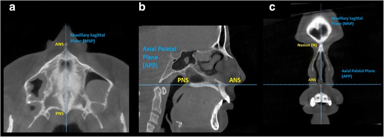
Mini-implant-assisted rapid palatal expansion (MARPE) appliances have been developed with the aim to enhance the orthopedic effect induced by rapid maxillary expansion (RME). Maxillary Skeletal Expander (MSE) is a particular type of MARPE appliance characterized by the presence of four mini-implants positioned in the posterior part of the palate with bi-cortical engagement. The aim of the present study is to evaluate the MSE effects on the midpalatal and pterygopalatine sutures in late adolescents, using high-resolution CBCT. Specific aims are to define the magnitude and sagittal parallelism of midpalatal suture opening, to measure the extent of transverse asymmetry of split, and to illustrate the possibility of splitting the pterygopalatine suture.

METHODS

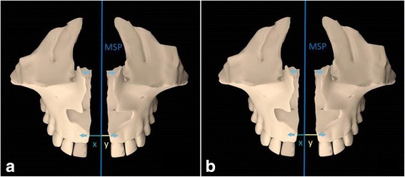
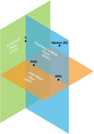
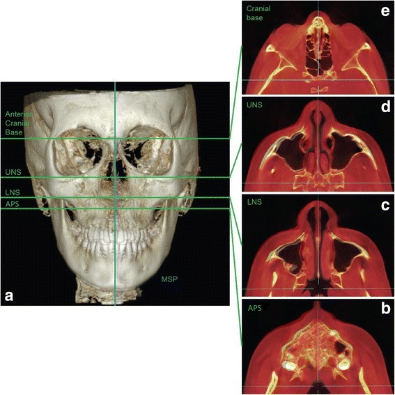
Fifteen subjects (mean age of 17.2 years; range, 13.9-26.2 years) were treated with MSE. Pre- and post-treatment CBCT exams were taken and superimposed. A novel methodology based on three new reference planes was utilized to analyze the sutural changes. Parameters were compared from pre- to post-treatment and between genders non-parametrically using the Wilcoxon sign rank test. For the frequency of openings in the lower part of the pterygopalatine suture, the Fisher's exact test was used.

RESULTS

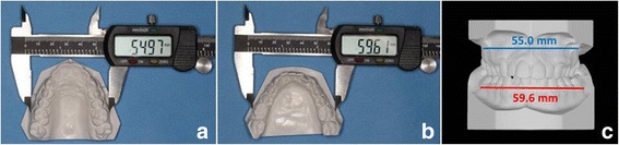
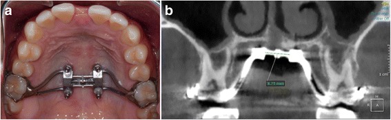
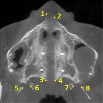
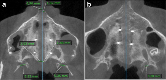
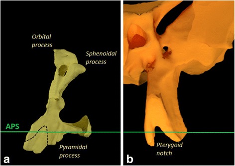
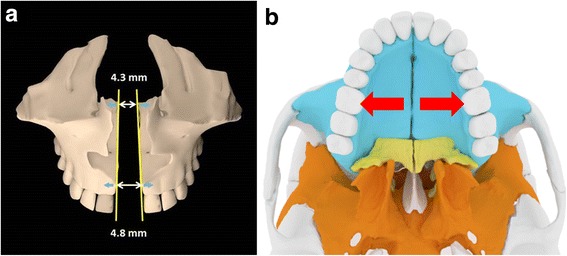
Regarding the magnitude of midpalatal suture opening, the split at anterior nasal spine (ANS) and at posterior nasal spine (PNS) was 4.8 and 4.3 mm, respectively. The amount of split at PNS was 90% of that at ANS, showing that the opening of the midpalatal suture was almost perfectly parallel antero-posteriorly. On average, one half of the anterior nasal spine (ANS) moved more than the contralateral one by 1.1 mm. Openings between the lateral and medial plates of the pterygoid process were detectable in 53% of the sutures (P < 0.05). No significant differences were found in the magnitude and frequency of suture opening between males and females. Correlation between age and suture opening was negligible (R range, 0.3-4.2%).

CONCLUSIONS

Midpalatal suture was successfully split by MSE in late adolescents, and the opening was almost perfectly parallel in a sagittal direction. Regarding the extent of transverse asymmetry of the split, on average one half of ANS moved more than the contralateral one by 1.1 mm. Pterygopalatine suture was split in its lower region by MSE, as the pyramidal process was pulled out from the pterygoid process. Patient gender and age had a negligible influence on suture opening for the age group considered in the study.

摘要

背景

微型植入物辅助快速腭扩张(MARPE)器具的开发旨在增强快速上颌扩张(RME)引起的矫形效果。上颌扩弓器(MSE)是一种特殊类型的 MARPE 器具,其特点是在后腭部有四个微型植入物,采用双皮质结合。本研究的目的是使用高分辨率 CBCT 评估 MARPE 对上颌中缝和翼腭缝的影响。具体目标是定义中缝张开的幅度和矢状平行度,测量分裂的横向不对称程度,并说明分裂翼腭缝的可能性。

方法

对 15 名受试者(平均年龄 17.2 岁;范围,13.9-26.2 岁)进行 MSE 治疗。拍摄治疗前后的 CBCT 检查并进行叠加。利用一种基于三个新参考平面的新方法来分析缝的变化。使用 Wilcoxon 符号秩检验对参数进行非参数比较,以比较治疗前后和性别之间的差异。对于翼腭缝下部开口的频率,使用 Fisher 确切检验。

结果

就中缝张开的幅度而言,前鼻棘(ANS)和后鼻棘(PNS)的分裂分别为 4.8 和 4.3mm。PNS 的分裂量为 ANS 的 90%,表明中缝的张开几乎是完全前后平行的。平均而言,一半的前鼻棘(ANS)比对侧移动 1.1mm。53%的翼状突外侧和内侧板之间有开口(P<0.05)。男性和女性之间在缝的张开幅度和频率上没有发现显著差异。年龄与缝张开之间的相关性可以忽略不计(R 范围,0.3-4.2%)。

结论

MSE 成功地在上颌骨晚期青少年中分裂了中缝,并且在矢状方向上的开口几乎是完全平行的。关于分裂的横向不对称程度,平均而言,一半的前鼻棘(ANS)比对侧移动 1.1mm。MSE 也将翼腭缝下部分开,因为尖牙从翼状突中被拔出。考虑到研究中的年龄组,患者性别和年龄对缝的张开影响可以忽略不计。

https://cdn.ncbi.nlm.nih.gov/pmc/blobs/dcdf/5663987/83bdae06cc9e/40510_2017_188_Fig11_HTML.jpg
https://cdn.ncbi.nlm.nih.gov/pmc/blobs/dcdf/5663987/518aa068d4f2/40510_2017_188_Fig1_HTML.jpg
https://cdn.ncbi.nlm.nih.gov/pmc/blobs/dcdf/5663987/014fd3fdeb16/40510_2017_188_Fig2_HTML.jpg
https://cdn.ncbi.nlm.nih.gov/pmc/blobs/dcdf/5663987/8679b97ec421/40510_2017_188_Fig3_HTML.jpg
https://cdn.ncbi.nlm.nih.gov/pmc/blobs/dcdf/5663987/169cf1254d30/40510_2017_188_Fig4_HTML.jpg
https://cdn.ncbi.nlm.nih.gov/pmc/blobs/dcdf/5663987/cf068f1b4fae/40510_2017_188_Fig5_HTML.jpg
https://cdn.ncbi.nlm.nih.gov/pmc/blobs/dcdf/5663987/ed76834fd0d6/40510_2017_188_Fig6_HTML.jpg
https://cdn.ncbi.nlm.nih.gov/pmc/blobs/dcdf/5663987/4ca476d45db4/40510_2017_188_Fig7_HTML.jpg
https://cdn.ncbi.nlm.nih.gov/pmc/blobs/dcdf/5663987/3115243a85d9/40510_2017_188_Fig8_HTML.jpg
https://cdn.ncbi.nlm.nih.gov/pmc/blobs/dcdf/5663987/ab01846515e2/40510_2017_188_Fig9_HTML.jpg
https://cdn.ncbi.nlm.nih.gov/pmc/blobs/dcdf/5663987/1b9bf948faf3/40510_2017_188_Fig10_HTML.jpg
https://cdn.ncbi.nlm.nih.gov/pmc/blobs/dcdf/5663987/83bdae06cc9e/40510_2017_188_Fig11_HTML.jpg

相似文献

1
Changes in the midpalatal and pterygopalatine sutures induced by micro-implant-supported skeletal expander, analyzed with a novel 3D method based on CBCT imaging.基于 CBCT 成像的新型 3D 方法分析微种植体支抗骨扩张器引起的中缝和翼腭缝变化。
Prog Orthod. 2017 Nov 1;18(1):34. doi: 10.1186/s40510-017-0188-7.
2
Tomographic assessment of palatal suture opening pattern and pterygopalatine suture disarticulation in the axial plane after midfacial skeletal expansion.面中部骨骼扩张后轴位上硬腭缝张开模式和翼腭缝分离的断层评估。
Prog Orthod. 2020 Jul 20;21(1):21. doi: 10.1186/s40510-020-00321-9.
3
Zygomaticomaxillary modifications in the horizontal plane induced by micro-implant-supported skeletal expander, analyzed with CBCT images.颧骨-上颌骨在微植体支抗骨扩张器作用下的水平向改建:CBCT 影像分析。
Prog Orthod. 2018 Oct 22;19(1):41. doi: 10.1186/s40510-018-0240-2.
4
Assessment of the bone thickness of the palate on cone-beam computed tomography for placement of miniscrew-assisted rapid palatal expansion appliances.评估应用于微型螺钉辅助快速腭扩张装置的腭部锥形束 CT 骨厚度。
Am J Orthod Dentofacial Orthop. 2022 Jun;161(6):849-857. doi: 10.1016/j.ajodo.2021.01.037. Epub 2022 Feb 9.
5
Dentoskeletal changes and their correlations after micro-implant-assisted palatal expansion (MARPE) in adults with advanced midpalatal suture ossification.成人硬腭缝骨化晚期行微种植体辅助腭扩张(MARPE)后的牙颌骨变化及其相关性。
Clin Oral Investig. 2022 Mar;26(3):3021-3031. doi: 10.1007/s00784-021-04284-x. Epub 2021 Nov 13.
6
Effects of bicortical anchorage on pterygopalatine suture opening with microimplant-assisted maxillary skeletal expansion.双皮质锚固对微种植体辅助上颌骨骨骼扩展时翼腭缝打开的影响。
Am J Orthod Dentofacial Orthop. 2021 Apr;159(4):502-511. doi: 10.1016/j.ajodo.2020.02.013. Epub 2021 Jan 22.
7
Age-dependent interactions of maxillary sutures during RME and their effects on palatal morphology : CBCT and dental cast analysis.上颌骨缝在扩弓治疗中的年龄相关性变化及其对上颌形态的影响:CBCT 和牙模分析。
J Orofac Orthop. 2022 Nov;83(6):412-431. doi: 10.1007/s00056-022-00429-z. Epub 2022 Oct 7.
8
Short-term effects of microimplant-assisted rapid palatal expansion on the circummaxillary sutures in skeletally mature patients: A cone-beam computed tomography study.微种植体辅助快速腭扩张对上颌周骨缝在成熟患者中的短期影响:锥形束 CT 研究。
Am J Orthod Dentofacial Orthop. 2022 Feb;161(2):e187-e197. doi: 10.1016/j.ajodo.2021.01.023. Epub 2021 Dec 3.
9
Midpalatal suture bone repair after miniscrew-assisted rapid palatal expansion in adults.成人微型骨钉辅助快速腭扩张后正中缝骨修复。
Prog Orthod. 2022 Oct 17;23(1):35. doi: 10.1186/s40510-022-00431-6.
10
Effects of bonded rapid palatal expansion on the transverse dimensions of the maxilla: a cone-beam computed tomography study.粘结式快速腭扩张对上颌横向尺寸的影响:锥形束 CT 研究。
Am J Orthod Dentofacial Orthop. 2010 Apr;137(4 Suppl):S79-85. doi: 10.1016/j.ajodo.2008.11.024.

引用本文的文献

1
Treatment outcomes of 3D-printed custom and conventional mini-implant assisted rapid palatal expanders (MARPE).3D打印定制式和传统微型种植体辅助快速腭扩展器(MARPE)的治疗效果
Prog Orthod. 2025 Aug 25;26(1):30. doi: 10.1186/s40510-025-00576-0.
2
Nasal and pharyngeal airway changes following three different rapid palatal expansion protocols in nongrowing subjects: Effects of miniscrew-assisted rapid palatal expansion and surgically-assisted rapid palatal expansion with or without pterygomaxillary disjunction.非生长发育期受试者采用三种不同的快速腭扩展方案后的鼻腔和咽气道变化:微螺钉辅助快速腭扩展以及有或无翼上颌裂离的外科辅助快速腭扩展的效果
Korean J Orthod. 2025 Jul 25;55(4):314-326. doi: 10.4041/kjod25.020. Epub 2025 May 7.
3

本文引用的文献

1
Effects of monocortical and bicortical mini-implant anchorage on bone-borne palatal expansion using finite element analysis.使用有限元分析研究单皮质和双皮质微型种植体支抗对骨支持式腭扩展的影响。
Am J Orthod Dentofacial Orthop. 2017 May;151(5):887-897. doi: 10.1016/j.ajodo.2016.10.025.
2
Soft tissue changes in the orofacial region after rapid maxillary expansion : A cone beam computed tomography study.快速上颌扩弓后口面部区域的软组织变化:一项锥形束计算机断层扫描研究。
J Orofac Orthop. 2017 May;78(3):193-200. doi: 10.1007/s00056-016-0074-9.
3
Microimplant-assisted rapid palatal expansion appliance to orthopedically correct transverse maxillary deficiency in an adult.
Mid-facial skeletal and soft tissue changes after maxillary skeletal expander application: a retrospective CBCT study.
上颌骨扩弓器应用后中面部骨骼和软组织的变化:一项回顾性CBCT研究
BMC Oral Health. 2025 Jul 15;25(1):1168. doi: 10.1186/s12903-025-06569-z.
4
[Structural changes in the craniofacial complex induced by microimplant-supported skeletal expander - MSE. a review].[微种植体支抗骨扩弓器-MSE诱导的颅面复合体结构变化。综述]
Rev Cient Odontol (Lima). 2025 May 16;13(2):e243. doi: 10.21142/2523-2754-1302-2025-243. eCollection 2025 Apr-Jun.
5
Correlation between and intraoral metal alloys.与口腔内金属合金之间的相关性。 (你提供的原文似乎不完整,“Correlation between”后面缺少具体内容)
Korean J Orthod. 2025 Jul 25;55(4):327-331. doi: 10.4041/kjod25.015. Epub 2025 Jun 2.
6
Comparison of Ultrasonography and Cone-beam Computed Tomography for Quantitative Assessment of Midpalatal Suture Opening after Rapid Palatal Expansion: A Pilot Study.超声检查与锥形束计算机断层扫描在快速扩弓后腭中缝开口定量评估中的比较:一项初步研究
J Med Ultrasound. 2024 Jun 30;33(1):54-60. doi: 10.4103/jmu.jmu_154_23. eCollection 2025 Jan-Mar.
7
A new removable connecting device for MARPE expander with non-common path of insertion.一种用于MARPE扩弓器的新型可移除连接装置,具有非共同插入路径。
Zhong Nan Da Xue Xue Bao Yi Xue Ban. 2024 Oct 28;49(10):1649-1662. doi: 10.11817/j.issn.1672-7347.2024.240283.
8
Comparison of Side Effects Between Miniscrew-Assisted Rapid Palatal Expansion (MARPE) and Surgically Assisted Rapid Palatal Expansion (SARPE) in Adult Patients: A Scoping Review.成人患者中微型螺钉辅助快速腭扩展(MARPE)与手术辅助快速腭扩展(SARPE)副作用的比较:一项范围综述
Dent J (Basel). 2025 Jan 22;13(2):47. doi: 10.3390/dj13020047.
9
Effect of miniscrew-assisted maxillary protraction on width changes in the circummaxillary sutures.微型螺钉辅助上颌骨前牵引对上颌周围缝宽度变化的影响。
Angle Orthod. 2025 May 1;95(3):283-289. doi: 10.2319/081424-665.1.
10
Is efficacy of miniscrew-assisted rapid palatal expansion in mid to late adolescents and young adults related to craniofacial sutures opening? A systematic review and meta-analysis.微型螺钉辅助快速腭扩展在青少年中后期及年轻成年人中的疗效与颅面骨缝开放有关吗?一项系统评价和荟萃分析。
Med Oral Patol Oral Cir Bucal. 2025 Mar 1;30(2):e271-e281. doi: 10.4317/medoral.26935.
微型种植体辅助快速腭扩展矫治器用于成人上颌横向发育不足的正畸矫治
Am J Orthod Dentofacial Orthop. 2016 May;149(5):716-28. doi: 10.1016/j.ajodo.2015.04.043.
4
Fast three-dimensional superimposition of cone beam computed tomography for orthopaedics and orthognathic surgery evaluation.用于骨科和正颌外科评估的锥形束计算机断层扫描快速三维叠加
Int J Oral Maxillofac Surg. 2015 Sep;44(9):1188-96. doi: 10.1016/j.ijom.2015.04.001. Epub 2015 Apr 29.
5
Cone-beam computed tomography evaluation of dentoskeletal changes after asymmetric rapid maxillary expansion.锥形束计算机断层扫描评估非对称快速上颌扩展后的牙颌骨骼变化。
Am J Orthod Dentofacial Orthop. 2015 Jan;147(1):61-71. doi: 10.1016/j.ajodo.2014.09.014.
6
Tooth-borne vs bone-borne rapid maxillary expanders in late adolescence.青春期后期牙支持式与骨支持式快速上颌扩弓器的比较
Angle Orthod. 2015 Mar;85(2):253-62. doi: 10.2319/030514-156.1. Epub 2014 Dec 9.
7
The effects of micro-implant assisted rapid palatal expansion (MARPE) on the nasomaxillary complex--a finite element method (FEM) analysis.微型种植体辅助快速腭扩展(MARPE)对鼻上颌复合体的影响——有限元法(FEM)分析
Prog Orthod. 2014 Aug 29;15(1):52. doi: 10.1186/s40510-014-0052-y.
8
An assessment of the maxilla after rapid maxillary expansion using cone beam computed tomography in growing children.使用锥形束计算机断层扫描对正在生长发育的儿童快速上颌扩弓后的上颌骨进行评估。
Dental Press J Orthod. 2014 Jan-Feb;19(1):26-35. doi: 10.1590/2176-9451.19.1.026-035.oar.
9
Effects of rapid maxillary expansion on the cranial and circummaxillary sutures.快速上颌扩展对颅和眶周缝的影响。
Am J Orthod Dentofacial Orthop. 2011 Oct;140(4):510-9. doi: 10.1016/j.ajodo.2010.10.024.
10
Immediate effects of rapid maxillary expansion with Haas-type and hyrax-type expanders: a randomized clinical trial.快速上颌扩张(哈斯式和赫克斯式扩弓器)即刻效果的随机临床试验。
Am J Orthod Dentofacial Orthop. 2011 Sep;140(3):366-76. doi: 10.1016/j.ajodo.2010.07.025.