Flasar Jan, Volk Gerd Fabian, Granitzka Thordis, Geißler Katharina, Irintchev Andrey, Lehmann Thomas, Guntinas-Lichius Orlando
Department of Otorhinolaryngology Jena University Hospital Jena Germany.
Facial Nerve Center Jena Jena University Hospital Jena Germany.
Laryngoscope Investig Otolaryngol. 2017 Sep 25;2(5):325-330. doi: 10.1002/lio2.95. eCollection 2017 Oct.
OBJECTIVES/HYPOTHESIS: The time course of the reinnervation of the paralyzed face after hypoglossal-facial jump nerve suture using electromyography (EMG) was assessed. The relation to the clinical outcome was analyzed.
Retrospective single-center cohort study.
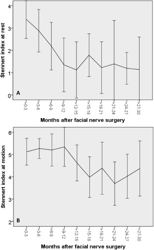
Reestablishment of motor units was studied by quantitative EMG and motor unit potential (MUP) analysis in 11 patients after hypoglossal-facial jump nerve suture. Functional recovery was evaluated using the Stennert index (0 = normal; 10 = maximal palsy).
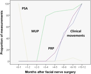
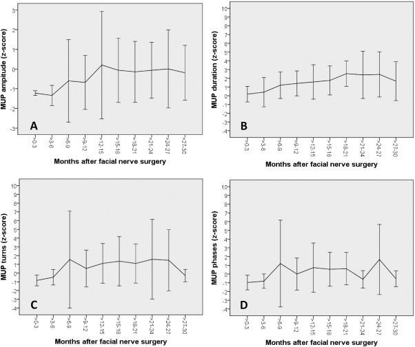
Clinically, first movements were seen between 6 and >10 months after surgery in individual patients. Maximal improvement was achieved at 18 months. The Stennert index decreased from 7.9 ± 2.0 preoperatively to a final postoperative score of 5.8 ± 2.4. EMG monitoring performed for 2.8 to 60 months after surgery revealed that pathological spontaneous activity disappeared within 2 weeks. MUPs were first recorded after the 2nd month and present in all 11 patients 8-10 months post-surgery. Polyphasic regeneration potentials first appeared at 4-10 months post-surgery. The MUP amplitudes increased between the 3rd and 15th months after surgery to values of control muscles. The MUP duration was significantly increased above normal values between the 3rd and 24th months after surgery.
Reinnervation can be detected at least 2 months earlier by EMG than by clinical evaluation. Changes should be followed for at least 18 months to assess outcome. EMG changes reflected the remodeling of motor units due to axonal regeneration and collateral sprouting by hypoglossal nerve fibers into the reinnervated facial muscle fibers.
3b.
目的/假设:采用肌电图(EMG)评估舌下-面神经跳跃神经缝合术后瘫痪面部再支配的时间进程,并分析其与临床结果的关系。
回顾性单中心队列研究。
对11例舌下-面神经跳跃神经缝合术后患者进行定量肌电图和运动单位电位(MUP)分析,研究运动单位的重建情况。使用施泰纳特指数(0=正常;10=最大麻痹)评估功能恢复情况。
临床上,个别患者在术后6至10个月以上出现首次运动。18个月时达到最大改善。施泰纳特指数从术前的7.9±2.0降至术后最终评分的5.8±2.4。术后2.8至60个月进行的肌电图监测显示,病理性自发电活动在2周内消失。术后第2个月后首次记录到运动单位电位,术后8至10个月所有11例患者均出现。多相再生电位在术后4至10个月首次出现。运动单位电位幅度在术后第3至15个月增加至对照肌肉的值。运动单位电位持续时间在术后第3至24个月显著高于正常值。
肌电图检测再支配比临床评估至少早2个月。应跟踪变化至少18个月以评估结果。肌电图变化反映了由于轴突再生和舌下神经纤维向再支配的面部肌肉纤维侧支发芽导致的运动单位重塑。
3b。