Kuroda Shintaro, Kobayashi Tsuyoshi, Ohdan Hideki
Department of Gastroenterological and Transplant Surgery, Hiroshima University Hospital, Hiroshima, Japan.
Department of Gastroenterological and Transplant Surgery, Hiroshima University Hospital, Hiroshima, Japan.
Int J Surg Case Rep. 2017;41:219-222. doi: 10.1016/j.ijscr.2017.10.015. Epub 2017 Oct 26.
The 3D printing model of the intrahepatic vessels and regional anatomy are often used for navigation surgery. Here, we report the use of the model for anatomical resection of hepatocellular carcinoma.
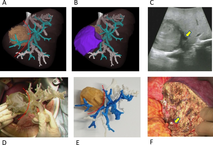
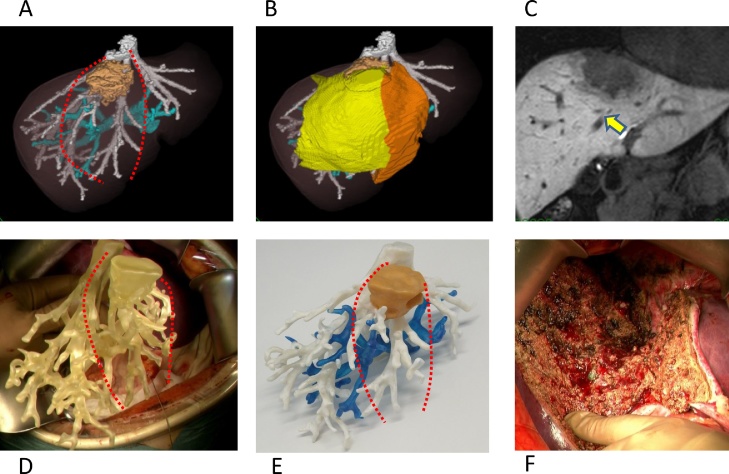
Case 1: A tumor, 31mm in diameter, was located in segment 7 of the liver. Using the 3D model, we identified the regional Glissonian pedicle and performed resection of segment 7. Case 2: The tumor was located in segment 4/8 and involved the middle hepatic vein. Radical resection of segment 4 and of the ventral area of the right anterior section was performed using the 3D model.
The positional relationship between the intrahepatic vessels and liver tumors is the most important factor for anatomical resection for hepatocellular carcinoma. Therefore our simplified 3D model of intrahepatic vessels without liver parenchyma is sufficient for effective guidance during surgery and has the advantage of being feasible to use for all HCC surgeries.
Use of 3D printed models might have many merits and contribute to the great improvement of the surgical quality.
肝内血管及区域解剖结构的3D打印模型常用于导航手术。在此,我们报告该模型在肝细胞癌解剖性切除术中的应用。
病例1:一个直径31mm的肿瘤位于肝脏第7段。利用3D模型,我们识别了区域肝蒂并进行了第7段切除。病例2:肿瘤位于第4/8段并累及肝中静脉。利用3D模型对第4段及右前叶腹侧区域进行了根治性切除。
肝内血管与肝肿瘤之间的位置关系是肝细胞癌解剖性切除的最重要因素。因此,我们简化的无肝实质的肝内血管3D模型足以在手术中提供有效指导,并且具有可用于所有肝癌手术的优势。
使用3D打印模型可能有诸多优点,并有助于手术质量的大幅提高。