Department of Pathology, Yong Loo Lin School of Medicine, National University of Singapore
Department of Pathology, National University Hospital, National University Health System, Singapore.
Haematologica. 2018 Feb;103(2):278-287. doi: 10.3324/haematol.2017.180430. Epub 2017 Nov 2.
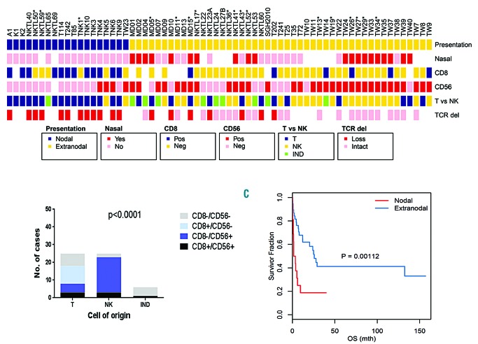
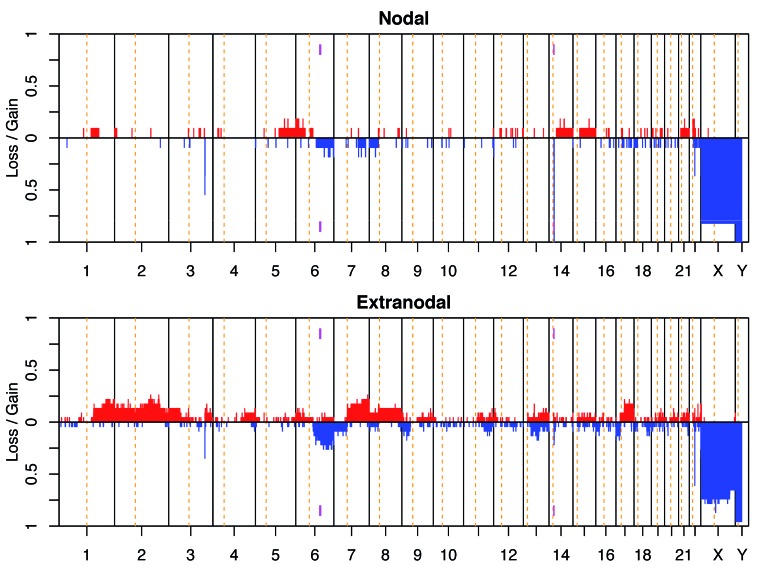
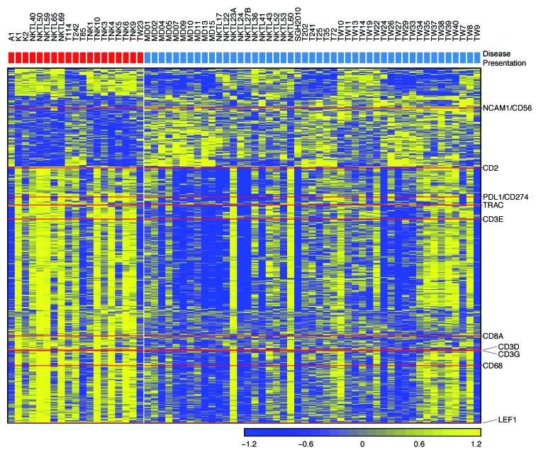
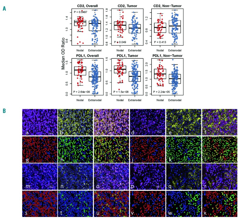
The molecular biology of primary nodal T- and NK-cell lymphoma and its relationship with extranodal NK/T-cell lymphoma, nasal type is poorly understood. In this study, we assessed the relationship between nodal and extranodal Epstein-Barr virus-positive T/NK-cell lymphomas using gene expression profiling and copy number aberration analyses. We performed gene expression profiling and copy number aberration analysis on 66 cases of Epstein-Barr virus-associated T/NK-cell lymphoma from nodal and extranodal sites, and correlated the molecular signatures with clinicopathological features. Three distinct molecular clusters were identified with one enriched for nodal presentation and loss of 14q11.2 (TCRA loci). T/NK-cell lymphomas with a nodal presentation (nodal-group) were significantly associated with older age, lack of nasal involvement, and T-cell lineage compared to those with an extranodal presentation (extranodal-group). On multivariate analysis, nodal presentation was an independent factor associated with short survival. Comparing the molecular signatures of the nodal and extranodal groups it was seen that the former was characterized by upregulation of PD-L1 and T-cell-related genes, including CD2 and CD8, and downregulation of CD56, consistent with the CD8/CD56-immunophenotype. PD-L1 and CD2 protein expression levels were validated using multiplexed immunofluorescence. Interestingly, nodal group lymphomas were associated with 14q11.2 loss which correlated with loss of TCR loci and T-cell origin. Overall, our results suggest that T/NK-cell lymphoma with nodal presentation is distinct and deserves to be classified separately from T/NK-cell lymphoma with extranodal presentation. Upregulation of PD-L1 indicates that it may be possible to use anti-PD1 immunotherapy in this distinctive entity. In addition, loss of 14q11.2 may be a potentially useful diagnostic marker of T-cell lineage.
原发性结内 T/NK 细胞淋巴瘤和其与结外 NK/T 细胞淋巴瘤,鼻型的分子生物学尚不清楚。在这项研究中,我们使用基因表达谱和拷贝数畸变分析来评估结内和结外 EBV 阳性 T/NK 细胞淋巴瘤之间的关系。我们对来自结内和结外部位的 66 例 EBV 相关 T/NK 细胞淋巴瘤进行了基因表达谱和拷贝数畸变分析,并将分子特征与临床病理特征相关联。我们鉴定出三个不同的分子簇,其中一个簇富集了结内表现和 14q11.2 缺失(TCR 基因座)。与结外表现(结外组)相比,具有结内表现(结内组)的 T/NK 细胞淋巴瘤与年龄较大、无鼻受累和 T 细胞谱系有关。在多变量分析中,结内表现是与生存时间短相关的独立因素。比较结内和结外组的分子特征,前者表现为 PD-L1 和 T 细胞相关基因(包括 CD2 和 CD8)的上调,以及 CD56 的下调,与 CD8/CD56-免疫表型一致。使用多重免疫荧光验证了 PD-L1 和 CD2 蛋白的表达水平。有趣的是,结内组淋巴瘤与 14q11.2 缺失相关,这与 TCR 基因座和 T 细胞起源的缺失相关。总的来说,我们的结果表明,具有结内表现的 T/NK 细胞淋巴瘤是独特的,值得与具有结外表现的 T/NK 细胞淋巴瘤分开分类。PD-L1 的上调表明,在这种独特的实体中,可能可以使用抗 PD1 免疫疗法。此外,14q11.2 的缺失可能是 T 细胞谱系的一个潜在有用的诊断标志物。