Tan Hongbao, Chen Li, Ma Jun
Department of Anesthesiology, Beijing Anzhen Hospital of Capital Medical University, Beijing 100029, P.R. China.
Department of Nephrology, The Second People's Hospital of Hunan, Changsha, Hunan 410007, P.R. China.
Exp Ther Med. 2017 Nov;14(5):4272-4278. doi: 10.3892/etm.2017.5089. Epub 2017 Sep 1.
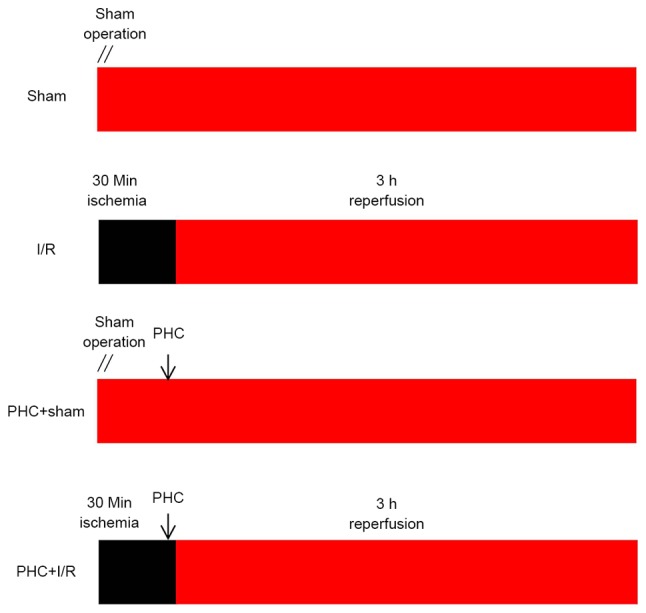
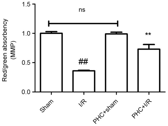
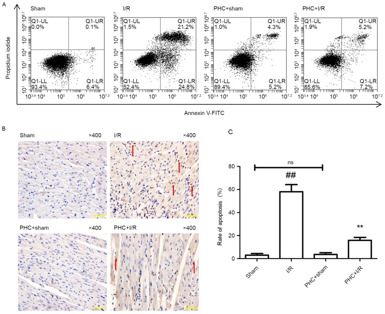
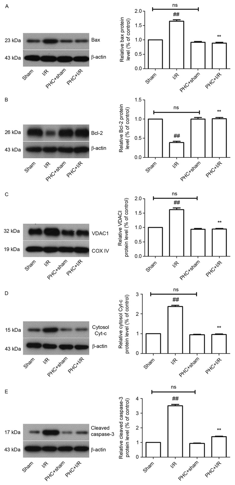
Ischemic heart disease is a major cause of mortality and disability worldwide. Timely reperfusion is currently the most effective method of treating ischemic heart disease; however, abrupt reperfusion may cause ischemia/reperfusion (I/R) injury. Apoptosis serves an important role in the progression of myocardial I/R injury and it has been demonstrated that the mitochondria are the center of regulation for apoptosis. Penehyclidine hydrochloride (PHC) is used during surgery and has recently been identified as a new type of anticholinergic drug. It has been demonstrated that pretreatment with PHC reduces myocardial apoptosis in rat hearts. The present study aimed to investigate the effects of PHC post-conditioning on myocardial cell apoptosis in a rat model of myocardial I/R and to determine whether the mitochondria-induced pathway was activated. Male Wistar rats were evenly and randomly categorized into 4 experimental groups as follows: i) Sham group; ii) I/R group; iii) PHC+sham group; and iv) PHC+I/R group. A PHC (1 mg/kg) post-conditioning approach (5 min before reperfusion) was used in addition to I/R in the PHC-treated groups. Following 3 h reperfusion, flow cytometry and terminal deoxynucleotidyl transferase dUTP nick end labeling staining were performed to measure myocardial cell apoptosis. A JC-1 staining method was performed to measure the mitochondrial membrane potential of myocardial cells. The expression of Bax, Bcl-2, voltage dependent anion-selective channel protein 1 (VDAC1), cytosol cytochrome (cyt-) and cleaved caspase-3 was analyzed using western blotting. PHC post-conditioning significantly reduced apoptosis in cardiomyocytes, significantly downregulated the expression of Bax, VDAC1, cytosol cytochrome and cleaved caspase-3 but significantly upregulated the expression of Bcl-2. PHC post-conditioning also restored the mitochondrial membrane potential. Thus, the present study demonstrated that PHC post-conditioning protects cardiomyocytes against apoptosis in the rat model of myocardial I/R by inhibiting the mitochondria-induced intrinsic pathway.
缺血性心脏病是全球范围内导致死亡和残疾的主要原因。及时再灌注是目前治疗缺血性心脏病最有效的方法;然而,突然再灌注可能会导致缺血/再灌注(I/R)损伤。细胞凋亡在心肌I/R损伤的进展中起重要作用,并且已经证明线粒体是细胞凋亡调控的中心。盐酸戊乙奎醚(PHC)在手术中使用,最近被确定为一种新型抗胆碱能药物。已经证明,用PHC预处理可减少大鼠心脏中的心肌细胞凋亡。本研究旨在探讨PHC后处理对心肌I/R大鼠模型心肌细胞凋亡的影响,并确定线粒体诱导途径是否被激活。雄性Wistar大鼠被均匀随机分为4个实验组,如下:i)假手术组;ii)I/R组;iii)PHC+假手术组;iv)PHC+I/R组。除了对PHC处理组进行I/R外,还采用了PHC(1mg/kg)后处理方法(再灌注前5分钟)。再灌注3小时后,进行流式细胞术和末端脱氧核苷酸转移酶dUTP缺口末端标记染色以测量心肌细胞凋亡。采用JC-1染色法测量心肌细胞的线粒体膜电位。使用蛋白质印迹法分析Bax、Bcl-2、电压依赖性阴离子选择性通道蛋白1(VDAC1)、细胞色素c(cyt-c)和裂解的caspase-3的表达。PHC后处理显著减少了心肌细胞凋亡,显著下调了Bax、VDAC1、细胞色素c和裂解的caspase-3的表达,但显著上调了Bcl-2的表达。PHC后处理还恢复了线粒体膜电位。因此,本研究表明,PHC后处理通过抑制线粒体诱导的内源性途径保护心肌I/R大鼠模型中的心肌细胞免于凋亡。