Department of Biomedical Sciences and Morphological and Functional Imaging, G. Martino University Hospital Messina, Via Consolare Valeria, 1, 98100, Messina, Italy.
Institute for Experimental and Translational Cardiovascular Imaging, DZHK Centre for Cardiovascular Imaging, University Hospital Frankfurt, Theodor-Stern- Kai 7, Frankfurt am Main, Germany.
J Cardiovasc Magn Reson. 2017 Nov 6;19(1):83. doi: 10.1186/s12968-017-0400-4.
Reducing time and contrast agent doses are important goals to provide cost-efficient cardiovascular magnetic resonance (CMR) imaging. Limited information is available regarding the feasibility of evaluating left ventricular (LV) function after gadobutrol injection as well as defining the lowest dose for high quality scar imaging. We sought to evaluate both aspects separately and systematically to provide an optimized protocol for contrast-enhanced CMR (CE-CMR) using gadobutrol.
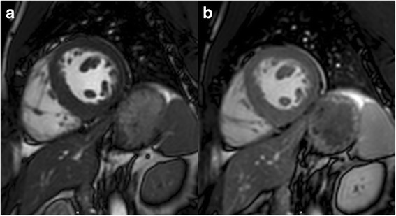
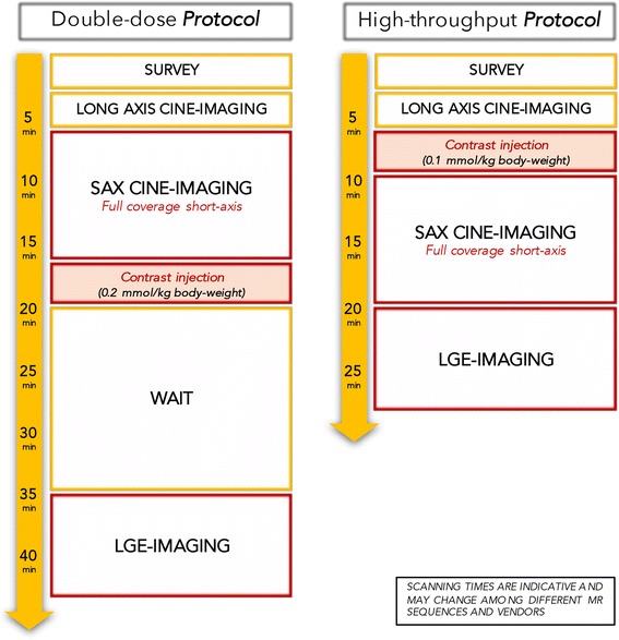
This is a prospective, randomized, single-blind cross-over study performed in two different populations. The first population consisted of 30 patients with general indications for a rest CE-CMR who underwent cine-imaging before and immediately after intravenous administration of 0.1 mmol/kg body-weight of gadobutrol. Quantitative assessment of LV volumes and function was performed by the same reader in a randomized and blinded fashion. The second population was composed of 30 patients with indication to late gadolinium enhancement (LGE) imaging, which was performed twice at different gadobutrol doses (0.1 mmol/kg vs. 0.2 mmol/kg) and at different time delays (5 and 10 min vs. 5, 10, 15 and 20 min), within a maximal interval of 21 days. LGE images were analysed qualitatively (contrast-to-noise ratio) and quantitatively (LGE%-of-mass).
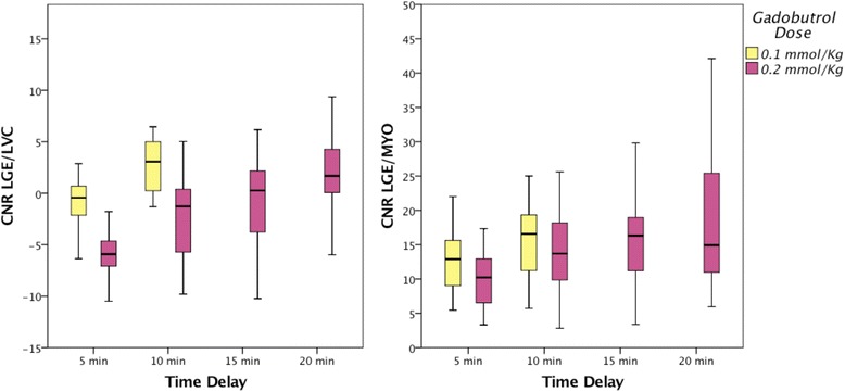
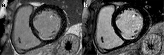
Excellent correlation between pre- and post-contrast cine-imaging was found, with no difference of LV stroke volume and ejection fraction (p = 0.538 and p = 0.095, respectively). End-diastolic-volume and end-systolic-volume were measured significantly larger after contrast injection (p = 0.008 and p = 0.001, respectively), with a mean difference of 3.7 ml and 2.9 ml, respectively. LGE imaging resulted in optimal contrast-to-noise ratios 10 min post-injection for a gadobutrol dose of 0.1 mmol/kg body-weight and 20 min for a dose of 0.2 mmol/kg body-weight. At these time points LGE quantification did not significantly differ (0.1 mmol/kg: 11% (16.4); 0.2 mmol/kg: 12% (14.5); p = 0.059), showing excellent correlation (ICC = 0.957; p < 0.001).
A standardized CE-CMR rest protocol giving a dose of 0.1 mmol/kg of gadobutrol before cine-imaging and performing LGE 10 min after injection represents a fast low-dose protocol without significant loss of information in comparison to a longer protocol with cine-imaging before contrast injection and a higher dose of gadobutrol. This approach allows to reduce examination time and costs as well as minimize contrast-agent exposure.
减少时间和造影剂剂量是提供具有成本效益的心血管磁共振(CMR)成像的重要目标。关于使用钆布醇注射后评估左心室(LV)功能以及确定高质量瘢痕成像的最低剂量的信息有限。我们旨在分别系统地评估这两个方面,为使用钆布醇的对比增强 CMR(CE-CMR)提供优化方案。
这是一项在两个不同人群中进行的前瞻性、随机、单盲交叉研究。第一组人群由 30 名具有一般静息 CE-CMR 适应证的患者组成,他们在静脉注射 0.1mmol/kg 体重的钆布醇前后立即进行电影成像。LV 容积和功能的定量评估由同一位读者以随机和盲法方式进行。第二组人群由 30 名具有延迟钆增强(LGE)成像适应证的患者组成,他们在 21 天的最大间隔内,以不同的钆布醇剂量(0.1mmol/kg 与 0.2mmol/kg)和不同的时间延迟(5 分钟与 10 分钟、5 分钟、10 分钟、15 分钟和 20 分钟)两次进行 LGE 成像。LGE 图像进行定性(对比噪声比)和定量(LGE%-of-mass)分析。
在电影成像前后发现了极好的相关性,LV 每搏量和射血分数没有差异(p=0.538 和 p=0.095)。注射造影剂后舒张末期容积和收缩末期容积明显增大(p=0.008 和 p=0.001),分别相差 3.7ml 和 2.9ml。对于 0.1mmol/kg 体重的剂量,LGE 成像在注射后 10 分钟获得最佳的对比噪声比,对于 0.2mmol/kg 体重的剂量则在 20 分钟获得最佳的对比噪声比。在这些时间点,LGE 定量没有显著差异(0.1mmol/kg:11%(16.4);0.2mmol/kg:12%(14.5);p=0.059),显示出极好的相关性(ICC=0.957;p<0.001)。
标准化的 CE-CMR 静息方案在电影成像前给予 0.1mmol/kg 的钆布醇剂量,并在注射后 10 分钟进行 LGE 成像,与在对比注射前进行更长时间的方案和更高剂量的钆布醇相比,是一种快速的低剂量方案,不会显著丢失信息。这种方法可以减少检查时间和成本,最大限度地减少造影剂暴露。