• 文献检索
  • 文档翻译
  • 深度研究
  • 学术资讯
  • Suppr Zotero 插件Zotero 插件
  • 邀请有礼
  • 套餐&价格
  • 历史记录
应用&插件
Suppr Zotero 插件Zotero 插件浏览器插件Mac 客户端Windows 客户端微信小程序
定价
高级版会员购买积分包购买API积分包
服务
文献检索文档翻译深度研究API 文档MCP 服务
关于我们
关于 Suppr公司介绍联系我们用户协议隐私条款
关注我们

Suppr 超能文献

核心技术专利:CN118964589B侵权必究
粤ICP备2023148730 号-1Suppr @ 2026

文献检索

告别复杂PubMed语法,用中文像聊天一样搜索,搜遍4000万医学文献。AI智能推荐,让科研检索更轻松。

立即免费搜索

文件翻译

保留排版,准确专业,支持PDF/Word/PPT等文件格式,支持 12+语言互译。

免费翻译文档

深度研究

AI帮你快速写综述,25分钟生成高质量综述,智能提取关键信息,辅助科研写作。

立即免费体验

缺血性慢性心肌病的多模态成像

Multimodality Imaging in Ischemic Chronic Cardiomyopathy.

作者信息

Muscogiuri Giuseppe, Guglielmo Marco, Serra Alessandra, Gatti Marco, Volpato Valentina, Schoepf Uwe Joseph, Saba Luca, Cau Riccardo, Faletti Riccardo, McGill Liam J, De Cecco Carlo Nicola, Pontone Gianluca, Dell'Aversana Serena, Sironi Sandro

机构信息

Department of Radiology, Istituto Auxologico Italiano IRCCS, San Luca Hospital, University Milano Bicocca, 20149 Milan, Italy.

Department of Cardiology, Division of Heart and Lungs, Utrecht University, Utrecht University Medical Center, 3584 Utrecht, The Netherlands.

出版信息

J Imaging. 2022 Feb 1;8(2):35. doi: 10.3390/jimaging8020035.

DOI:10.3390/jimaging8020035
PMID:35200737
原文链接:https://pmc.ncbi.nlm.nih.gov/articles/PMC8877428/
Abstract

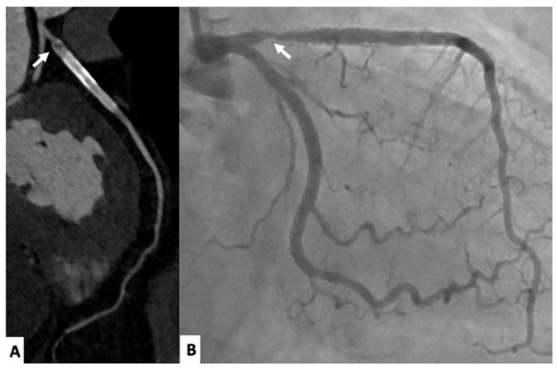
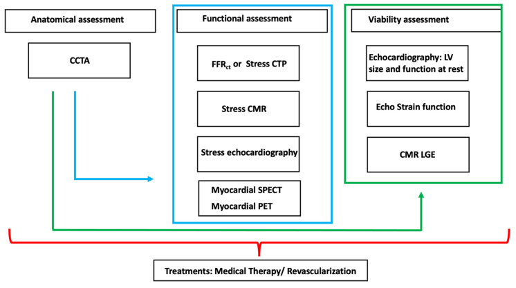
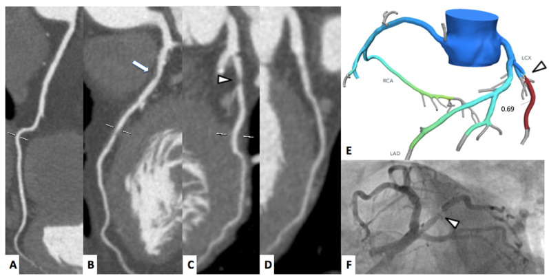
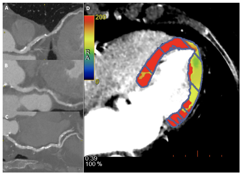
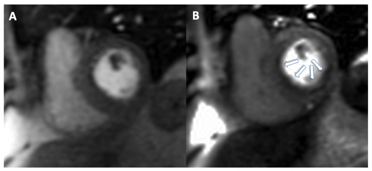
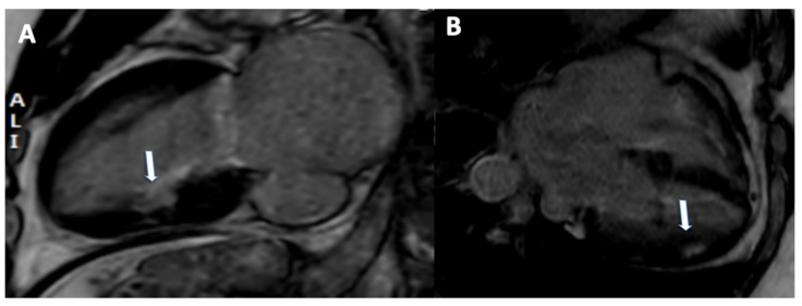
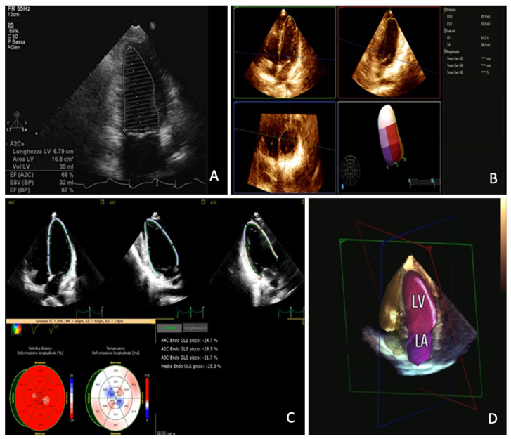
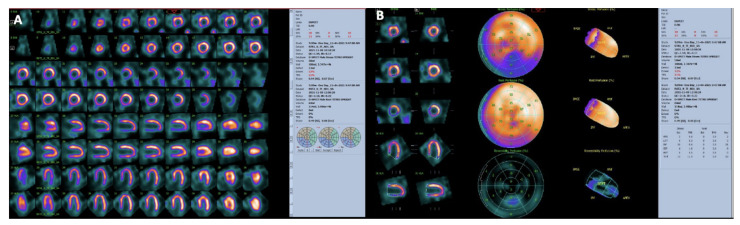
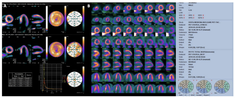
Ischemic chronic cardiomyopathy (ICC) is still one of the most common cardiac diseases leading to the development of myocardial ischemia, infarction, or heart failure. The application of several imaging modalities can provide information regarding coronary anatomy, coronary artery disease, myocardial ischemia and tissue characterization. In particular, coronary computed tomography angiography (CCTA) can provide information regarding coronary plaque stenosis, its composition, and the possible evaluation of myocardial ischemia using fractional flow reserve CT or CT perfusion. Cardiac magnetic resonance (CMR) can be used to evaluate cardiac function as well as the presence of ischemia. In addition, CMR can be used to characterize the myocardial tissue of hibernated or infarcted myocardium. Echocardiography is the most widely used technique to achieve information regarding function and myocardial wall motion abnormalities during myocardial ischemia. Nuclear medicine can be used to evaluate perfusion in both qualitative and quantitative assessment. In this review we aim to provide an overview regarding the different noninvasive imaging techniques for the evaluation of ICC, providing information ranging from the anatomical assessment of coronary artery arteries to the assessment of ischemic myocardium and myocardial infarction. In particular this review is going to show the different noninvasive approaches based on the specific clinical history of patients with ICC.

摘要

缺血性慢性心肌病(ICC)仍然是导致心肌缺血、梗死或心力衰竭的最常见心脏疾病之一。多种成像方式的应用可以提供有关冠状动脉解剖结构、冠状动脉疾病、心肌缺血和组织特征的信息。特别是,冠状动脉计算机断层扫描血管造影(CCTA)可以提供有关冠状动脉斑块狭窄、其成分的信息,以及使用血流储备分数CT或CT灌注对心肌缺血进行可能的评估。心脏磁共振成像(CMR)可用于评估心脏功能以及缺血的存在。此外,CMR可用于表征冬眠或梗死心肌的心肌组织。超声心动图是获取心肌缺血期间心脏功能和心肌壁运动异常信息最广泛使用的技术。核医学可用于定性和定量评估灌注。在本综述中,我们旨在概述用于评估ICC的不同无创成像技术,提供从冠状动脉解剖评估到缺血心肌和心肌梗死评估的信息。特别是,本综述将展示基于ICC患者特定临床病史的不同无创方法。

https://cdn.ncbi.nlm.nih.gov/pmc/blobs/e1df/8877428/9904144d9b6b/jimaging-08-00035-g009.jpg
https://cdn.ncbi.nlm.nih.gov/pmc/blobs/e1df/8877428/84977b3c9e2a/jimaging-08-00035-g001.jpg
https://cdn.ncbi.nlm.nih.gov/pmc/blobs/e1df/8877428/7c9ec909183d/jimaging-08-00035-g002.jpg
https://cdn.ncbi.nlm.nih.gov/pmc/blobs/e1df/8877428/65698c9285a9/jimaging-08-00035-g003.jpg
https://cdn.ncbi.nlm.nih.gov/pmc/blobs/e1df/8877428/63c48a1f9a79/jimaging-08-00035-g004.jpg
https://cdn.ncbi.nlm.nih.gov/pmc/blobs/e1df/8877428/bbbb92764183/jimaging-08-00035-g005.jpg
https://cdn.ncbi.nlm.nih.gov/pmc/blobs/e1df/8877428/50e6b0057ef0/jimaging-08-00035-g006.jpg
https://cdn.ncbi.nlm.nih.gov/pmc/blobs/e1df/8877428/37100c1b1627/jimaging-08-00035-g007.jpg
https://cdn.ncbi.nlm.nih.gov/pmc/blobs/e1df/8877428/39e17898306e/jimaging-08-00035-g008.jpg
https://cdn.ncbi.nlm.nih.gov/pmc/blobs/e1df/8877428/9904144d9b6b/jimaging-08-00035-g009.jpg

相似文献

1
Multimodality Imaging in Ischemic Chronic Cardiomyopathy.缺血性慢性心肌病的多模态成像
J Imaging. 2022 Feb 1;8(2):35. doi: 10.3390/jimaging8020035.
2
Coronary computed tomography angiography (CCTA) and cardiac magnetic resonance (CMR) imaging in the assessment of patients presenting with chest pain suspected for acute coronary syndrome.冠状动脉计算机断层扫描血管造影术(CCTA)和心脏磁共振成像(CMR)在评估因疑似急性冠状动脉综合征而出现胸痛的患者中的应用。
Ann Transl Med. 2016 Jul;4(13):255. doi: 10.21037/atm.2016.06.30.
3
Positron emission tomography for the assessment of myocardial viability: an evidence-based analysis.用于评估心肌活力的正电子发射断层扫描:一项基于证据的分析。
Ont Health Technol Assess Ser. 2005;5(16):1-167. Epub 2005 Oct 1.
4
Multimodality imaging evaluation of Chagas disease: an expert consensus of Brazilian Cardiovascular Imaging Department (DIC) and the European Association of Cardiovascular Imaging (EACVI).恰加斯病的多模态成像评估:巴西心血管成像部(DIC)与欧洲心血管成像协会(EACVI)的专家共识
Eur Heart J Cardiovasc Imaging. 2018 Apr 1;19(4):459-460n. doi: 10.1093/ehjci/jex154.
5
Roles of nuclear cardiology, cardiac computed tomography, and cardiac magnetic resonance: assessment of patients with suspected coronary artery disease.核素心脏病学、心脏计算机断层扫描和心脏磁共振成像的作用:疑似冠状动脉疾病患者的评估
J Nucl Med. 2006 Jan;47(1):74-82.
6
[Noninvasive diagnostic of coronary artery disease].[冠状动脉疾病的无创诊断]
Ther Umsch. 2009 Apr;66(4):241-51. doi: 10.1024/0040-5930.66.4.241.
7
CAD detection in patients with intermediate-high pre-test probability: low-dose CT delayed enhancement detects ischemic myocardial scar with moderate accuracy but does not improve performance of a stress-rest CT perfusion protocol.在具有中高度预检测概率的患者中进行 CAD 检测:低剂量 CT 延迟增强以中等准确度检测缺血性心肌瘢痕,但不能提高负荷-静息 CT 灌注方案的性能。
JACC Cardiovasc Imaging. 2013 Oct;6(10):1062-1071. doi: 10.1016/j.jcmg.2013.04.013. Epub 2013 Sep 4.
8
Validation of dynamic 3-dimensional whole heart magnetic resonance myocardial perfusion imaging against fractional flow reserve for the detection of significant coronary artery disease.动态 3 维全心脏磁共振心肌灌注成像与血流储备分数检测用于诊断显著冠状动脉疾病的验证。
J Am Coll Cardiol. 2012 Aug 21;60(8):756-65. doi: 10.1016/j.jacc.2012.02.075. Epub 2012 Jul 18.
9
Noninvasive assessment of coronary anatomy and myocardial perfusion: going toward an integrated imaging approach.冠状动脉解剖结构和心肌灌注的无创评估:迈向综合成像方法
J Cardiovasc Med (Hagerstown). 2008 Oct;9(10):977-86. doi: 10.2459/JCM.0b013e328306f311.
10
Single photon emission computed tomography for the diagnosis of coronary artery disease: an evidence-based analysis.单光子发射计算机断层扫描用于冠状动脉疾病的诊断:一项基于证据的分析。
Ont Health Technol Assess Ser. 2010;10(8):1-64. Epub 2010 Jun 1.

引用本文的文献

1
Characterization of Coronary Artery Disease in Sepsis Survivors.脓毒症幸存者冠状动脉疾病的特征
Biomedicines. 2025 May 13;13(5):1181. doi: 10.3390/biomedicines13051181.
2
Interlinking pathways: a narrative review on the role of IL-6 in cancer and atherosclerosis.相互关联的通路:关于白细胞介素-6在癌症和动脉粥样硬化中作用的叙述性综述
Cardiovasc Diagn Ther. 2024 Dec 31;14(6):1186-1201. doi: 10.21037/cdt-24-344. Epub 2024 Nov 12.
3
Clinical Application of Myocardial Perfusion SPECT in Patients with Suspected or Known Coronary Artery Disease. What Role in the Multimodality Imaging Era?

本文引用的文献

1
Cardiac Magnetic Resonance Radiomics Reveal Differential Impact of Sex, Age, and Vascular Risk Factors on Cardiac Structure and Myocardial Tissue.心脏磁共振影像组学揭示性别、年龄和血管危险因素对心脏结构和心肌组织的不同影响。
Front Cardiovasc Med. 2021 Dec 22;8:763361. doi: 10.3389/fcvm.2021.763361. eCollection 2021.
2
2021 AHA/ACC/ASE/CHEST/SAEM/SCCT/SCMR Guideline for the Evaluation and Diagnosis of Chest Pain: A Report of the American College of Cardiology/American Heart Association Joint Committee on Clinical Practice Guidelines.2021 AHA/ACC/ASE/CHEST/SAEM/SCCT/SCMR 胸痛评估与诊断指南:美国心脏病学会/美国心脏协会联合临床实践指南委员会的报告。
J Am Coll Cardiol. 2021 Nov 30;78(22):e187-e285. doi: 10.1016/j.jacc.2021.07.053. Epub 2021 Oct 28.
3
心肌灌注单光子发射计算机断层扫描在疑似或已知冠状动脉疾病患者中的临床应用。在多模态成像时代发挥着什么作用?
Rev Cardiovasc Med. 2023 Feb 6;24(2):48. doi: 10.31083/j.rcm2402048. eCollection 2023 Feb.
4
Atrial and Ventricular Strain Imaging Using CMR in the Prediction of Ventricular Arrhythmia in Patients with Myocarditis.使用心脏磁共振成像进行心房和心室应变成像在预测心肌炎患者室性心律失常中的应用
J Clin Med. 2024 Jan 23;13(3):662. doi: 10.3390/jcm13030662.
5
The Role of Non-Invasive Multimodality Imaging in Chronic Coronary Syndrome: Anatomical and Functional Pathways.非侵入性多模态成像在慢性冠状动脉综合征中的作用:解剖学和功能途径
Diagnostics (Basel). 2023 Jun 16;13(12):2083. doi: 10.3390/diagnostics13122083.
6
[Prediction of significant coronary lesions by SPECT myocardial perfusion. Results from a national reference hospital in Lima-Peru].[通过单光子发射计算机断层扫描心肌灌注预测显著冠状动脉病变。秘鲁利马一家国家参考医院的结果]
Arch Peru Cardiol Cir Cardiovasc. 2022 Jun 27;3(2):74-81. doi: 10.47487/apcyccv.v3i2.222. eCollection 2022 Apr-Jun.
7
Editorial: Multimodality imaging in the assessment of ischemic chronic coronary syndrome.社论:缺血性慢性冠状动脉综合征评估中的多模态成像
Front Cardiovasc Med. 2023 Apr 11;10:1146050. doi: 10.3389/fcvm.2023.1146050. eCollection 2023.
8
The chronic coronary syndrome-Heart failure roundabout: A multimodality imaging workflow approach.慢性冠状动脉综合征-心力衰竭的循环路径:一种多模态成像工作流程方法。
Front Cardiovasc Med. 2022 Nov 22;9:1019529. doi: 10.3389/fcvm.2022.1019529. eCollection 2022.
9
Application of AI in cardiovascular multimodality imaging.人工智能在心血管多模态成像中的应用。
Heliyon. 2022 Oct 5;8(10):e10872. doi: 10.1016/j.heliyon.2022.e10872. eCollection 2022 Oct.
10
Multimodality Imaging of Sudden Cardiac Death and Acute Complications in Acute Coronary Syndrome.急性冠状动脉综合征中心脏性猝死及急性并发症的多模态成像
J Clin Med. 2022 Sep 26;11(19):5663. doi: 10.3390/jcm11195663.
CAD-RADS may underestimate coronary plaque progression as detected by serial CT angiography.CAD-RADS 可能低估了通过连续 CT 血管造影检测到的冠状动脉斑块进展。
Eur Heart J Cardiovasc Imaging. 2022 Oct 20;23(11):1530-1539. doi: 10.1093/ehjci/jeab215.
4
Prognostic value of coronary computed tomography angiographic derived fractional flow reserve: a systematic review and meta-analysis.基于冠状动脉 CT 血管造影的血流储备分数的预后价值:系统评价和荟萃分析。
Heart. 2022 Feb;108(3):194-202. doi: 10.1136/heartjnl-2021-319773. Epub 2021 Oct 22.
5
Artificial Intelligence Based Multimodality Imaging: A New Frontier in Coronary Artery Disease Management.基于人工智能的多模态成像:冠状动脉疾病管理的新前沿。
Front Cardiovasc Med. 2021 Sep 22;8:736223. doi: 10.3389/fcvm.2021.736223. eCollection 2021.
6
Coronary plaque features on CTA can identify patients at increased risk of cardiovascular events.CTA 上的冠状动脉斑块特征可识别心血管事件风险增加的患者。
Curr Opin Cardiol. 2021 Nov 1;36(6):784-792. doi: 10.1097/HCO.0000000000000917.
7
Challenges and opportunities to delivering cardiac imaging training: a national survey by the Italian college of cardiac radiology.提供心脏成像培训的挑战与机遇:意大利心脏放射学会的一项全国性调查
Insights Imaging. 2021 Sep 27;12(1):136. doi: 10.1186/s13244-021-01076-5.
8
Cardiac Magnetic Resonance Tissue Characterization in Ischemic Cardiomyopathy.缺血性心肌病的心脏磁共振组织特征。
J Thorac Imaging. 2022 Jan 1;37(1):2-16. doi: 10.1097/RTI.0000000000000621.
9
Feasibility of late gadolinium enhancement (LGE) in ischemic cardiomyopathy using 2D-multisegment LGE combined with artificial intelligence reconstruction deep learning noise reduction algorithm.基于 2D 多节段钆延迟增强(LGE)联合人工智能重建深度学习降噪算法评估缺血性心肌病 LGE 的可行性。
Int J Cardiol. 2021 Nov 15;343:164-170. doi: 10.1016/j.ijcard.2021.09.012. Epub 2021 Sep 10.
10
Coronary computed tomography angiography in patients with stable coronary artery disease.稳定性冠心病患者的冠状动脉 CT 血管造影。
Trends Cardiovasc Med. 2022 Oct;32(7):421-428. doi: 10.1016/j.tcm.2021.08.009. Epub 2021 Aug 25.