Department of Medical Imaging and Intervention, Imaging Core Laboratory, Institute for Radiological Research, Chang Gung Memorial Hospital at Linkou and Chang Gung University, Taoyuan, Taiwan.
Clinical Phenome Center, Chang Gung Memorial Hospital at Linkou, Taoyuan, Taiwan.
Contrast Media Mol Imaging. 2017 Oct 9;2017:6053879. doi: 10.1155/2017/6053879. eCollection 2017.
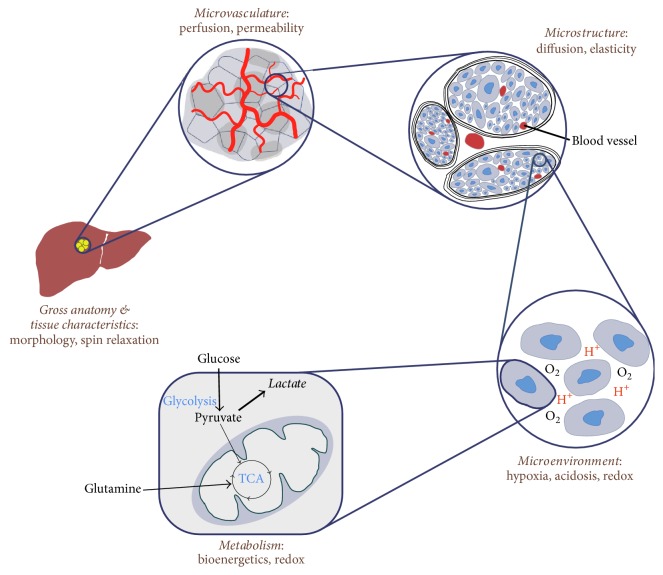
Cancer cells reprogram their metabolism to maintain viability via genetic mutations and epigenetic alterations, expressing overall dynamic heterogeneity. The complex relaxation mechanisms of nuclear spins provide unique and convertible tissue contrasts, making magnetic resonance imaging (MRI) and magnetic resonance spectroscopy (MRS) pertinent imaging tools in both clinics and research. In this review, we summarized MR methods that visualize tumor characteristics and its metabolic phenotypes on an anatomical, microvascular, microstructural, microenvironmental, and metabolomics scale. The review will progress from the utilities of basic spin-relaxation contrasts in cancer imaging to more advanced imaging methods that measure tumor-distinctive parameters such as perfusion, water diffusion, magnetic susceptibility, oxygenation, acidosis, redox state, and cell death. Analytical methods to assess tumor heterogeneity are also reviewed in brief. Although the clinical utility of tumor heterogeneity from imaging is debatable, the quantification of tumor heterogeneity using functional and metabolic MR images with development of robust analytical methods and improved MR methods may offer more critical roles of tumor heterogeneity data in clinics. MRI/MRS can also provide insightful information on pharmacometabolomics, biomarker discovery, disease diagnosis and prognosis, and treatment response. With these future directions in mind, we anticipate the widespread utilization of these MR-based techniques in studying cancer biology to better address significant clinical needs.
癌细胞通过基因突变和表观遗传改变重新编程其代谢以维持生存能力,表现出整体动态异质性。核自旋的复杂弛豫机制提供了独特且可转换的组织对比,使磁共振成像(MRI)和磁共振波谱(MRS)成为临床和研究中相关的成像工具。在这篇综述中,我们总结了在解剖学、微血管、微结构、微环境和代谢组学尺度上可视化肿瘤特征及其代谢表型的磁共振方法。本综述将从癌症成像中基本自旋弛豫对比的应用进展到更先进的成像方法,这些方法可以测量肿瘤特有的参数,如灌注、水扩散、磁化率、氧合、酸中毒、氧化还原状态和细胞死亡。我们还简要回顾了评估肿瘤异质性的分析方法。尽管成像中肿瘤异质性的临床应用存在争议,但使用功能和代谢 MRI 图像对肿瘤异质性进行定量,结合强大的分析方法和改进的 MRI 方法的发展,可能会为肿瘤异质性数据在临床中的应用提供更关键的作用。MRI/MRS 还可以提供关于药效代谢组学、生物标志物发现、疾病诊断和预后以及治疗反应的有见地的信息。考虑到这些未来的方向,我们预计这些基于磁共振的技术将在癌症生物学研究中得到广泛应用,以更好地满足重大临床需求。