Nezafat Maryam, Ramos Isabel T, Henningsson Markus, Protti Andrea, Basha Tamer, Botnar René M
Division of Imaging Sciences & Biomedical Engineering, King's College London, London, United Kingdom.
Department of Medicine, Beth Israel Deaconess Medical Center and Harvard Medical School, Boston, Massachusetts, United States of America.
PLoS One. 2017 Nov 9;12(11):e0187621. doi: 10.1371/journal.pone.0187621. eCollection 2017.
To develop and evaluate a 2D modified Look-Locker (MOLLI) for high-resolution T1 mapping in mice using a 3T MRI scanner.
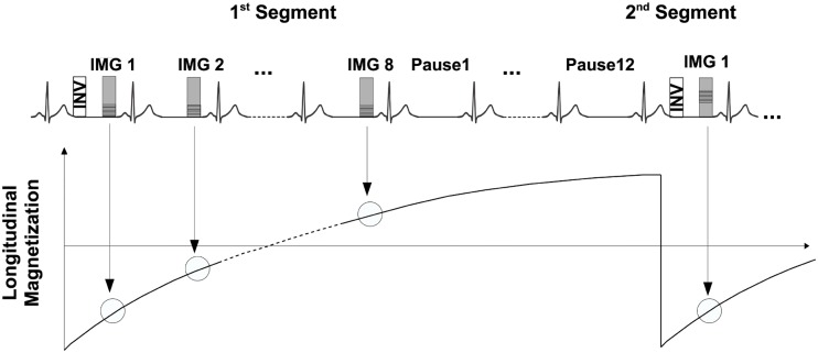
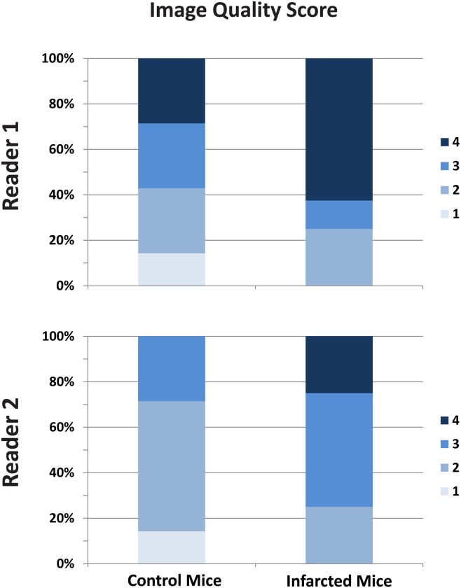
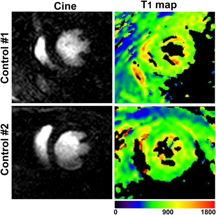
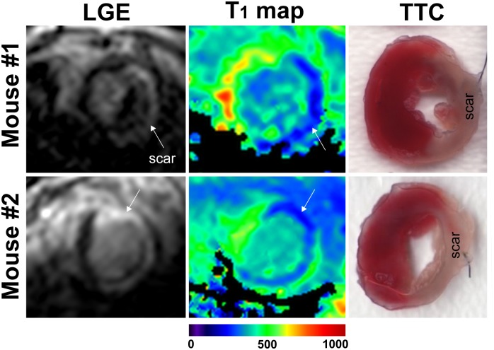
To allow high-resolution T1 mapping in mice at high heart rates a multi-shot ECG-triggered 2D MOLLI sequence was developed. In the proposed T1 mapping sequence the optimal number of sampling points and pause cardiac cycles following an initial adiabatic inversion pulse was investigated in a phantom. Seven native control and eight mice, 3 days post myocardial infarction (MI) after administration of gadolinium were scanned. Two experienced readers graded the visual T1 map quality.
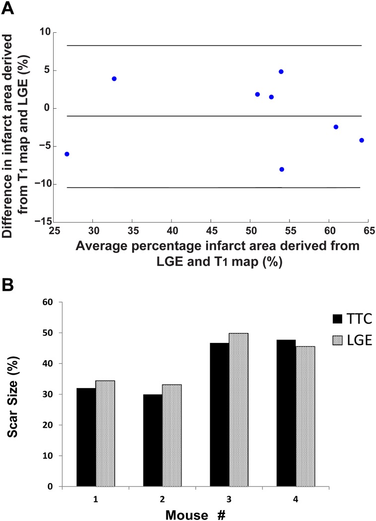
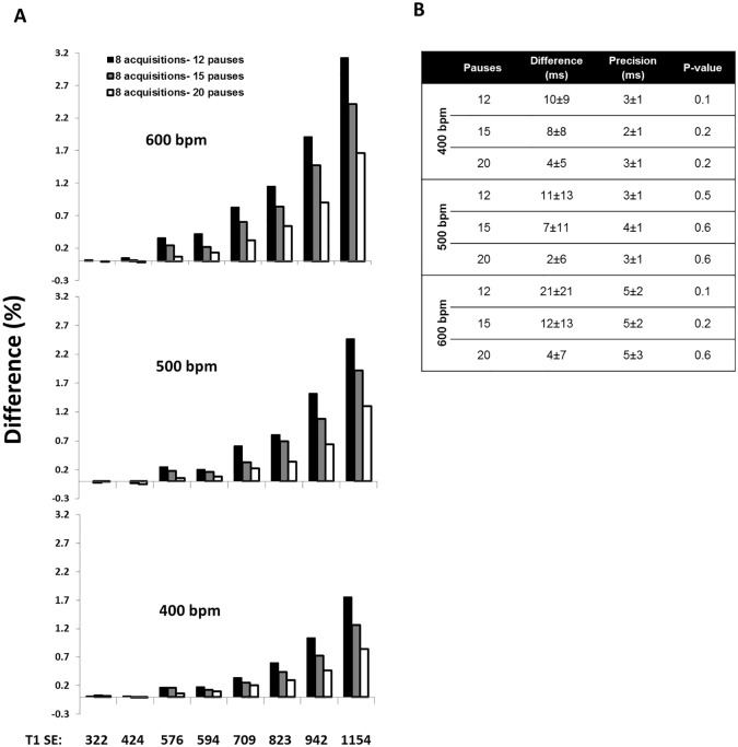
In T1 phantoms, there were no significant differences (<0.4% error) between 12, 15 and 20 pause cardiac cycles (p = 0.1, 0.2 and 0.6 respectively) for 8 acquisition cardiac cycles for 600bpm in comparison to the conventional inversion recovery spin echo T1 mapping sequence for short T1's (<600 ms). Subsequently, all in-vivo scans were performed with 8 data acquisitions and 12 pause cardiac cycles to minimize scan time. The mean native T1 value of myocardium in control animal was 820.5±52 ms. The post-contrast T1 measured 3 days after MI in scar was 264±59 ms and in healthy myocardium was 512±62 ms. The Bland-Altman analysis revealed mean difference of only -1.06% of infarct size percentage between T1 maps and LGE.
A multi-shot 2D MOLLI sequence has been presented that allows reliable measurement of high spatial resolution T1 maps in mice for heart rates up to 600bpm.
利用3T磁共振成像(MRI)扫描仪开发并评估一种用于小鼠高分辨率T1映射的二维改良Look-Locker(MOLLI)序列。
为了在高心率下对小鼠进行高分辨率T1映射,开发了一种多激发心电图触发的二维MOLLI序列。在所提出的T1映射序列中,在体模中研究了初始绝热反转脉冲后的最佳采样点数和暂停心动周期数。对7只正常对照小鼠和8只在给予钆后3天发生心肌梗死(MI)的小鼠进行扫描。两名经验丰富的阅片者对T1映射图的视觉质量进行评分。
在T1体模中,对于600次/分钟的心率,8个采集心动周期下,12、15和20个暂停心动周期之间无显著差异(误差<0.4%)(p分别为0.1、0.2和0.6),与传统反转恢复自旋回波T1映射序列相比,短T1值(<600毫秒)时情况相同。随后,所有体内扫描均采用8次数据采集和12个暂停心动周期,以尽量缩短扫描时间。对照动物心肌的平均固有T1值为820.5±52毫秒。MI后3天在瘢痕中的对比后T1值为264±59毫秒,在健康心肌中为512±62毫秒。Bland-Altman分析显示,T1映射图与延迟强化成像(LGE)之间梗死面积百分比的平均差异仅为-1.06%。
提出了一种多激发二维MOLLI序列,该序列能够在心率高达600次/分钟的小鼠中可靠地测量高空间分辨率的T1映射图。