Ishikawa Shohei, Noma Takahisa, Fu Hai Ying, Matsuzaki Takashi, Ishizawa Makoto, Ishikawa Kaori, Murakami Kazushi, Nishimoto Naoki, Nishiyama Akira, Minamino Tetsuo
Department of Cardiorenal and Cerebrovascular Medicine, Faculty of Medicine, Kagawa University, Kagawa, Japan.
Department of Cardiovascular Medicine, Osaka University Graduate School of Medicine, Suita, Osaka, Japan.
PLoS One. 2017 Nov 9;12(11):e0187894. doi: 10.1371/journal.pone.0187894. eCollection 2017.
Cardiac rupture is an important cause of death in the acute phase after myocardial infarction (MI). Macrophages play a pivotal role in cardiac remodeling after MI. Apoptosis inhibitor of macrophage (AIM) is secreted specifically by macrophages and contributes to macrophage accumulation in inflamed tissue by maintaining survival and recruiting macrophages. In this study, we evaluated the role of AIM in macrophage accumulation in the infarcted myocardium and cardiac rupture after MI.
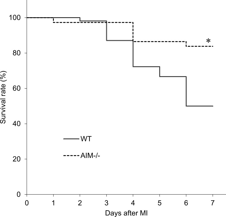
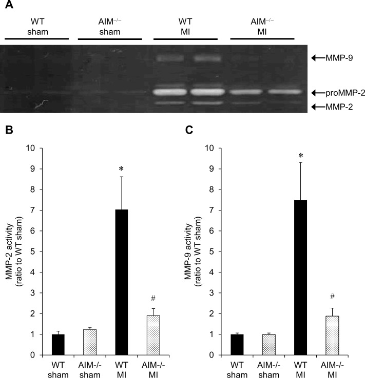
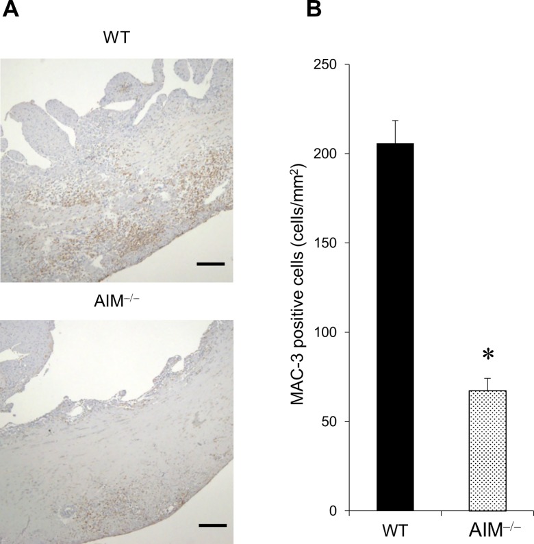
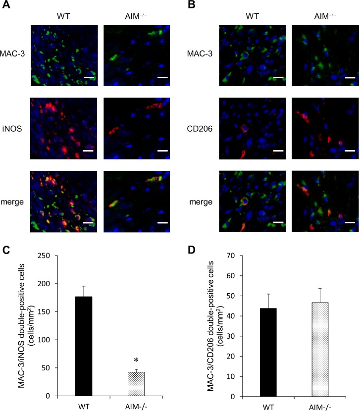
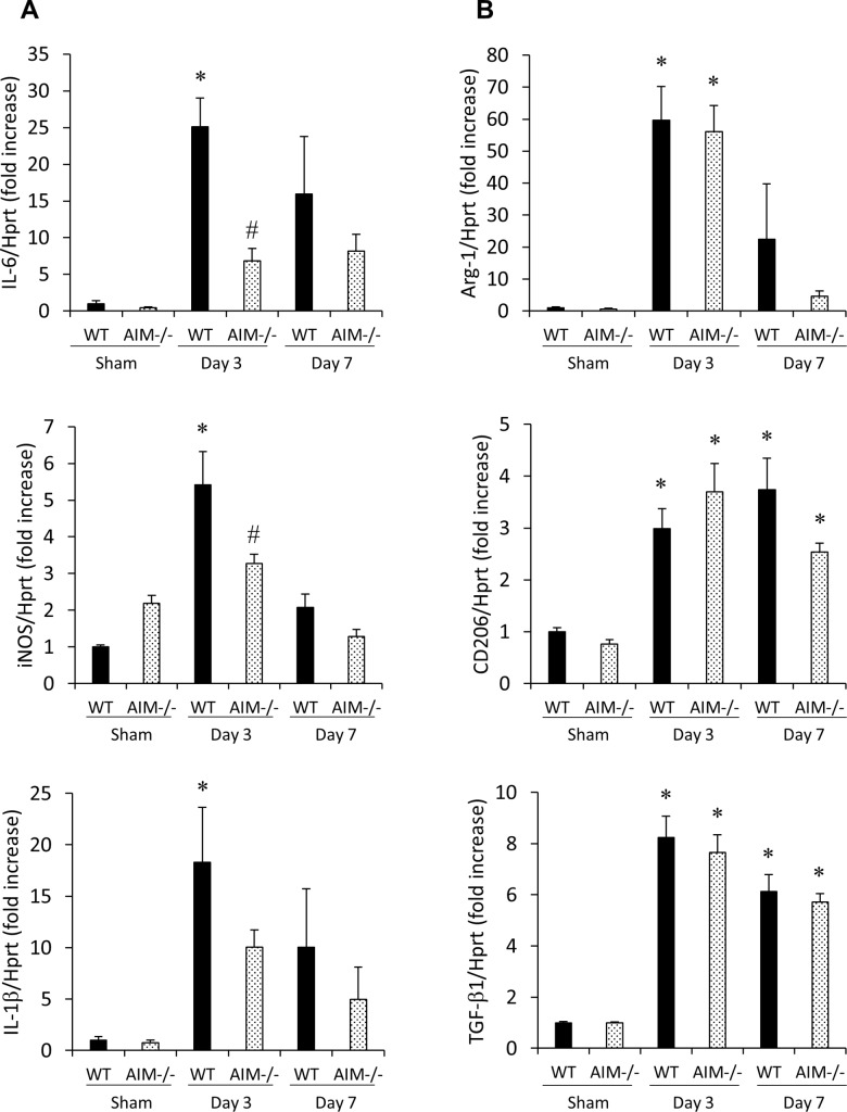
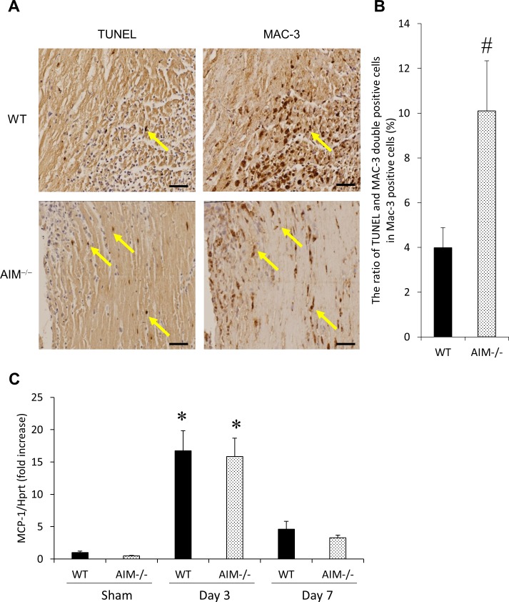
Wild-type (WT) and AIM‒/‒ mice underwent permanent left coronary artery ligation and were followed-up for 7 days. Macrophage accumulation and phenotypes (M1 pro-inflammatory macrophage or M2 anti-inflammatory macrophage) were evaluated by immunohistological analysis and RT-PCR. Matrix metalloproteinase (MMP) activity levels were measured by gelatin zymography. The survival rate was significantly higher (81.1% vs. 48.2%, P<0.05), and the cardiac rupture rate was significantly lower in AIM‒/‒ mice than in WT mice (10.8% vs. 31.5%, P<0.05). The number of M1 macrophages and the expression levels of M1 markers (iNOS and IL-6) in the infarcted myocardium were significantly lower in AIM‒/‒ mice than in WT mice. In contrast, there was no difference in the number of M2 macrophages and the expression of M2 markers (Arg-1, CD206 and TGF-β1) between the two groups. The ratio of apoptotic macrophages in the total macrophages was significantly higher in AIM‒/‒ mice than in WT mice, although MCP-1 expression did not differ between the two groups. MMP-2 and 9 activity levels in the infarcted myocardium were significantly lower in AIM‒/‒ mice than in WT mice.
These findings suggest that AIM depletion decreases the levels of M1 macrophages, which are a potent source of MMP-2 and 9, in the infarcted myocardium in the acute phase after MI by promoting macrophage apoptosis, and leads to a decrease in the incidence of cardiac rupture and improvements in survival rates.
心脏破裂是心肌梗死(MI)急性期死亡的重要原因。巨噬细胞在心肌梗死后的心脏重塑中起关键作用。巨噬细胞凋亡抑制因子(AIM)由巨噬细胞特异性分泌,通过维持存活和募集巨噬细胞促进巨噬细胞在炎症组织中的积聚。在本研究中,我们评估了AIM在心肌梗死后梗死心肌中巨噬细胞积聚和心脏破裂中的作用。
野生型(WT)和AIM基因敲除(AIM‒/‒)小鼠接受永久性左冠状动脉结扎,并随访7天。通过免疫组织学分析和逆转录-聚合酶链反应(RT-PCR)评估巨噬细胞积聚和表型(M1促炎巨噬细胞或M2抗炎巨噬细胞)。通过明胶酶谱法测量基质金属蛋白酶(MMP)活性水平。AIM‒/‒小鼠的存活率显著更高(81.1%对48.2%,P<0.05),且心脏破裂率显著低于WT小鼠(10.8%对31.5%,P<0.05)。AIM‒/‒小鼠梗死心肌中M1巨噬细胞数量和M1标志物(诱导型一氧化氮合酶(iNOS)和白细胞介素-6(IL-6))的表达水平显著低于WT小鼠。相反,两组之间M2巨噬细胞数量和M2标志物(精氨酸酶-1(Arg-1)、CD206和转化生长因子-β1(TGF-β1))的表达无差异。尽管两组之间单核细胞趋化蛋白-1(MCP-1)表达无差异,但AIM‒/‒小鼠中凋亡巨噬细胞在总巨噬细胞中的比例显著高于WT小鼠。AIM‒/‒小鼠梗死心肌中MMP-2和9的活性水平显著低于WT小鼠。
这些发现表明,AIM缺失通过促进巨噬细胞凋亡降低了心肌梗死后急性期梗死心肌中M1巨噬细胞的水平,而M1巨噬细胞是MMP-2和9的重要来源,并导致心脏破裂发生率降低和存活率提高。