Xu Yanyan, Yamashiro Tsuneo, Moriya Hiroshi, Tsubakimoto Maho, Tsuchiya Nanae, Nagatani Yukihiro, Matsuoka Shin, Murayama Sadayuki
Department of Radiology, Graduate School of Medical Science, University of the Ryukyus, Nishihara, Japan.
Department of Radiology, China-Japan Friendship Hospital, Beijing, People's Republic of China.
Int J Chron Obstruct Pulmon Dis. 2017 Oct 26;12:3123-3131. doi: 10.2147/COPD.S145599. eCollection 2017.
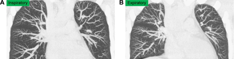
The aims of this study were to evaluate dynamic changes in heart size during the respiratory cycle using four-dimensional computed tomography (CT) and to understand the relationship of these changes to airflow limitation in smokers.
A total of 31 smokers, including 13 with COPD, underwent four-dimensional dynamic-ventilation CT during regular breathing. CT data were continuously reconstructed every 0.5 s, including maximum cross-sectional area (CSA) of the heart and mean lung density (MLD). Concordance between the cardiac CSA and MLD time curves was expressed by cross-correlation coefficients. The CT-based cardiothoracic ratio at inspiration and expiration was also calculated. Comparisons of the CT indices between COPD patients and non-COPD smokers were made using the Mann-Whitney test. Spearman rank correlation analysis was used to evaluate associations between CT indices and the forced expiratory volume in 1 s (FEV) relative to the forced vital capacity (FVC).
Cardiac CSA at both inspiration and expiration was significantly smaller in COPD patients than in non-COPD smokers (<0.05). The cross-correlation coefficient between cardiac CSA and MLD during expiration significantly correlated with FEV/FVC (ρ=0.63, <0.001), suggesting that heart size decreases during expiration in COPD patients. The change in the cardiothoracic ratio between inspiration and expiration frames was significantly smaller in COPD patients than in non-COPD smokers (<0.01).
Patients with COPD have smaller heart size on dynamic-ventilation CT than non-COPD smokers and have abnormal cardiac compression during expiration.
本研究旨在使用四维计算机断层扫描(CT)评估呼吸周期中心脏大小的动态变化,并了解这些变化与吸烟者气流受限之间的关系。
共有31名吸烟者,包括13名慢性阻塞性肺疾病(COPD)患者,在正常呼吸期间接受了四维动态通气CT检查。CT数据每0.5秒连续重建一次,包括心脏的最大横截面积(CSA)和平均肺密度(MLD)。心脏CSA和MLD时间曲线之间的一致性用互相关系数表示。还计算了吸气和呼气时基于CT的心胸比率。使用Mann-Whitney检验比较COPD患者和非COPD吸烟者之间的CT指标。采用Spearman等级相关分析评估CT指标与1秒用力呼气量(FEV)相对于用力肺活量(FVC)之间的关联。
COPD患者吸气和呼气时的心脏CSA均显著小于非COPD吸烟者(<0.05)。呼气时心脏CSA与MLD之间的互相关系数与FEV/FVC显著相关(ρ=0.63,<0.001),表明COPD患者呼气时心脏大小减小。COPD患者吸气和呼气帧之间心胸比率的变化显著小于非COPD吸烟者(<0.01)。
与非COPD吸烟者相比,COPD患者在动态通气CT上心脏较小,呼气时存在异常心脏受压。