Müller Elisabeth, Christopoulos Panagiotis F, Halder Sanjib, Lunde Anna, Beraki Kahsai, Speth Martin, Øynebråten Inger, Corthay Alexandre
Tumor Immunology Lab, Department of Pathology, Rikshospitalet, Oslo University Hospital, University of Oslo, Oslo, Norway.
Department of Biosciences, University of Oslo, Oslo, Norway.
Front Immunol. 2017 Oct 26;8:1383. doi: 10.3389/fimmu.2017.01383. eCollection 2017.
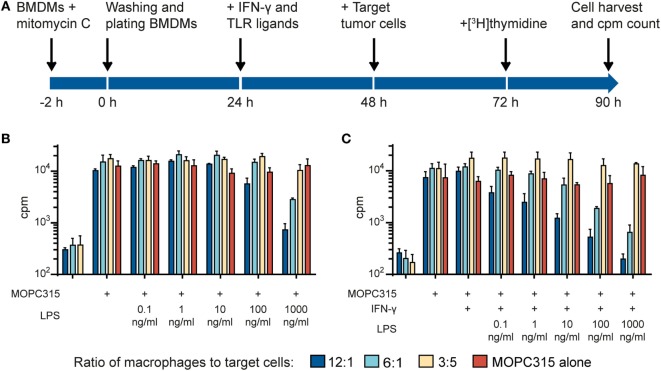
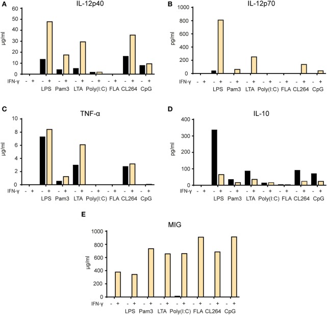
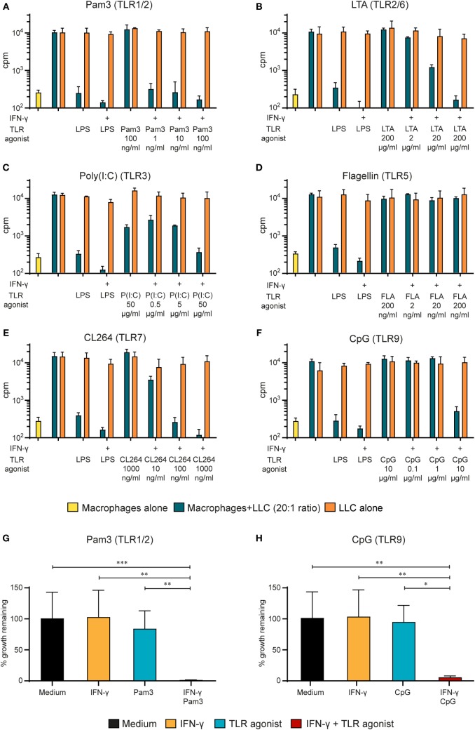
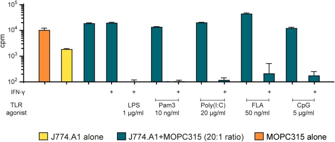
Tumor-associated macrophages may either promote or suppress tumor growth depending on their activation status. Interferon-γ (IFN-γ) has been identified as a key factor for inducing tumoricidal M1 phenotype in macrophages. However, it remains unclear whether IFN-γ is sufficient or if additional stimuli are required. Here, we tested IFN-γ and a panel of toll-like receptor (TLR) agonists for the ability to activate murine macrophages toward a tumoricidal M1 phenotype. The following TLR ligands were used: TLR1/TLR2 agonist Pam3CSK4, TLR2/TLR6 agonist lipotechoic acid, TLR3 agonist poly(I:C), TLR4 agonist lipopolysaccharide (LPS), TLR5 agonist flagellin, TLR7 agonist CL264, and TLR9 agonist CpG. We used an growth inhibition assay to measure both cytotoxic and cytostatic activity of mouse macrophages against Lewis lung carcinoma (LLC) and MOPC315 plasmacytoma tumor cells. Production of nitric oxide (NO) and cytokines by activated macrophages was quantified. We found that IFN-γ alone was not able to render macrophages tumoricidal. Similarly, macrophage activation with single TLR agonists was inefficient. In sharp contrast, IFN-γ was shown to synergize with TLR agonists for induction of macrophage tumoricidal activity and production of both NO and pro-inflammatory cytokines (TNF-α, IL-12p40, and IL-12p70). Furthermore, IFN-γ was shown to suppress macrophage IL-10 secretion induced by TLR agonists. NO production was necessary for macrophage tumoricidal activity. We conclude that two signals from the microenvironment are required for optimal induction of antitumor M1 macrophage phenotype. Combination treatment with IFN-γ and TLR agonists may offer new avenues for macrophage-based cancer immunotherapy.
肿瘤相关巨噬细胞根据其激活状态,可能促进或抑制肿瘤生长。干扰素-γ(IFN-γ)已被确定为诱导巨噬细胞产生杀肿瘤M1表型的关键因素。然而,尚不清楚IFN-γ是否足够,或者是否需要其他刺激因素。在此,我们测试了IFN-γ和一组Toll样受体(TLR)激动剂激活小鼠巨噬细胞向杀肿瘤M1表型转变的能力。使用了以下TLR配体:TLR1/TLR2激动剂Pam3CSK4、TLR2/TLR6激动剂脂磷壁酸、TLR3激动剂聚肌苷酸-聚胞苷酸(poly(I:C))、TLR4激动剂脂多糖(LPS)、TLR5激动剂鞭毛蛋白、TLR7激动剂CL264和TLR9激动剂CpG。我们使用生长抑制试验来测量小鼠巨噬细胞对刘易斯肺癌(LLC)和MOPC315浆细胞瘤肿瘤细胞的细胞毒性和细胞增殖抑制活性。对活化巨噬细胞产生的一氧化氮(NO)和细胞因子进行了定量分析。我们发现单独的IFN-γ不能使巨噬细胞具有杀肿瘤活性。同样,用单一TLR激动剂激活巨噬细胞效率低下。形成鲜明对比的是,IFN-γ与TLR激动剂协同作用可诱导巨噬细胞杀肿瘤活性以及产生NO和促炎细胞因子(肿瘤坏死因子-α、白细胞介素-12p40和白细胞介素-12p70)。此外,IFN-γ可抑制TLR激动剂诱导的巨噬细胞白细胞介素-10分泌。NO的产生对于巨噬细胞的杀肿瘤活性是必需的。我们得出结论,微环境中的两个信号是最佳诱导抗肿瘤M1巨噬细胞表型所必需的。IFN-γ和TLR激动剂联合治疗可能为基于巨噬细胞的癌症免疫治疗提供新途径。