Weinberg Marc S, Shachar Shlomit S, Muss Hyman B, Deal Allison M, Popuri Karteek, Yu Hyeon, Nyrop Kirsten A, Alston Shani M, Williams Grant R
School of Medicine, University of North Carolina at Chapel Hill, Chapel Hill, NC, USA.
Division of Oncology, Rambam Health Care Campus, Haifa, Israel.
Breast J. 2018 May;24(3):278-284. doi: 10.1111/tbj.12952. Epub 2017 Nov 15.
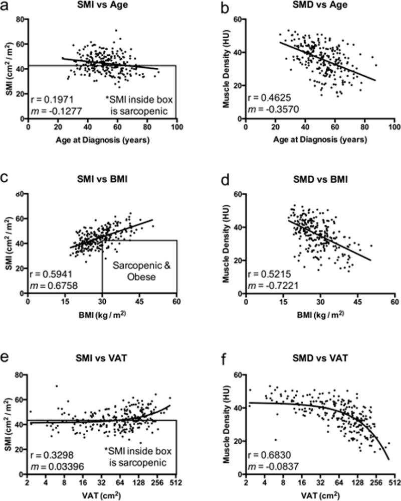
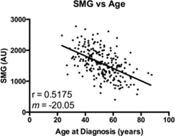
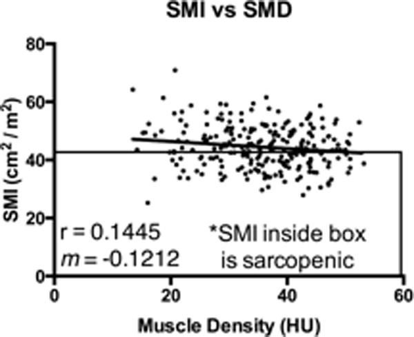
Skeletal muscle loss, commonly known as sarcopenia, is highly prevalent and prognostic of adverse outcomes in oncology. However, there is limited information on adults with early breast cancer and examination of other skeletal muscle indices, despite the potential prognostic importance. This study characterizes and examines age-related changes in body composition of adults with early breast cancer and describes the creation of a novel integrated muscle measure. Female patients diagnosed with stage I-III breast cancer with abdominal computerized tomography (CT) scans within 12 weeks from diagnosis were identified from local tumor registry (N = 241). Skeletal muscle index (muscle area per height [cm /m ]), skeletal muscle density, and subcutaneous and visceral adipose tissue areas, were determined from CT L3 lumbar segments. We calculated a novel integrated skeletal measure, skeletal muscle gauge, which combines skeletal muscle index and density (SMI × SMD). 241 patients were identified with available CT imaging. Median age 52 years and range of 23-87. Skeletal muscle index and density significantly decreased with age. Using literature based cut-points, older adults (≥65 years) had significantly higher proportions of sarcopenia (63 vs 28%) and myosteatosis (90 vs 11%) compared to younger adults (<50 years). Body mass index was positively correlated with skeletal muscle index and negatively correlated with muscle density. Skeletal muscle gauge correlated better with increasing age (ρ = 0.52) than with either skeletal muscle index (ρ = 0.20) or density (ρ = 0.46). Wide variations and age-related changes in body composition metrics were found using routinely obtained abdominal CT imaging. Skeletal muscle index and density provide independent, complementary information, and the product of the two metrics, skeletal muscle gauge, requires further research to explore its impact on outcomes in women with curable breast cancer.
骨骼肌丢失,通常称为肌肉减少症,在肿瘤学中非常普遍且是不良预后的指标。然而,尽管早期乳腺癌成年患者的其他骨骼肌指标可能具有预后重要性,但关于这方面的信息却很有限。本研究对早期乳腺癌成年患者身体成分的年龄相关变化进行了特征描述和分析,并介绍了一种新的综合肌肉测量方法的创建。从当地肿瘤登记处识别出在诊断后12周内进行腹部计算机断层扫描(CT)的I-III期乳腺癌女性患者(N = 241)。从CT扫描的L3腰椎节段确定骨骼肌指数(每身高[cm²/m²]的肌肉面积)、骨骼肌密度以及皮下和内脏脂肪组织面积。我们计算了一种新的综合骨骼测量指标——骨骼肌量规,它结合了骨骼肌指数和密度(SMI×SMD)。共识别出241例有可用CT影像的患者。中位年龄52岁,年龄范围为23 - 87岁。骨骼肌指数和密度随年龄显著下降。使用基于文献的切点,与年轻成年人(<50岁)相比,老年人(≥65岁)的肌肉减少症(63%对28%)和肌少脂症(90%对11%)比例显著更高。体重指数与骨骼肌指数呈正相关,与肌肉密度呈负相关。骨骼肌量规与年龄增长的相关性(ρ = 0.52)比与骨骼肌指数(ρ = 0.20)或密度(ρ = 0.46)的相关性更好。使用常规获得的腹部CT影像发现身体成分指标存在广泛差异和年龄相关变化。骨骼肌指数和密度提供独立的、互补的信息,这两个指标的乘积——骨骼肌量规,需要进一步研究以探讨其对可治愈乳腺癌女性预后的影响。