• 文献检索
  • 文档翻译
  • 深度研究
  • 学术资讯
  • Suppr Zotero 插件Zotero 插件
  • 邀请有礼
  • 套餐&价格
  • 历史记录
应用&插件
Suppr Zotero 插件Zotero 插件浏览器插件Mac 客户端Windows 客户端微信小程序
定价
高级版会员购买积分包购买API积分包
服务
文献检索文档翻译深度研究API 文档MCP 服务
关于我们
关于 Suppr公司介绍联系我们用户协议隐私条款
关注我们

Suppr 超能文献

核心技术专利:CN118964589B侵权必究
粤ICP备2023148730 号-1Suppr @ 2026

文献检索

告别复杂PubMed语法,用中文像聊天一样搜索,搜遍4000万医学文献。AI智能推荐,让科研检索更轻松。

立即免费搜索

文件翻译

保留排版,准确专业,支持PDF/Word/PPT等文件格式,支持 12+语言互译。

免费翻译文档

深度研究

AI帮你快速写综述,25分钟生成高质量综述,智能提取关键信息,辅助科研写作。

立即免费体验

基于临床和影像学特征的联合方法,用于区分非肿瘤性与肿瘤性垂体柄病变。

Joint approach based on clinical and imaging features to distinguish non-neoplastic from neoplastic pituitary stalk lesions.

作者信息

Lee Ji Ye, Park Ji Eun, Shim Woo Hyun, Jung Seung Chai, Choi Choong Gon, Kim Sang Joon, Kim Jeong Hoon, Kim Ho Sung

机构信息

Department of Radiology, Soonchunhyang University Bucheon Hospital, 170 Jomaru-ro, Wonmi-gu, Bucheon, Korea.

Department of Radiology and Research Institute of Radiology, University of Ulsan College of Medicine, Asan Medical Center, Seoul, Korea.

出版信息

PLoS One. 2017 Nov 15;12(11):e0187989. doi: 10.1371/journal.pone.0187989. eCollection 2017.

DOI:10.1371/journal.pone.0187989
PMID:29140989
原文链接:https://pmc.ncbi.nlm.nih.gov/articles/PMC5687750/
Abstract

PURPOSE

Distinguishing non-neoplastic pituitary stalk lesions (non-NPSLs) from neoplastic pituitary stalk lesions (NPSLs) is a major concern in guiding treatment for a thickened pituitary stalk. Our study aimed to aid provide preoperative diagnostic assistance by combining clinical and magnetic resonance imaging (MRI) findings to distinguish non-NPSLs from NPSLs.

MATERIALS AND METHODS

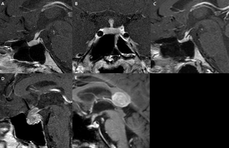
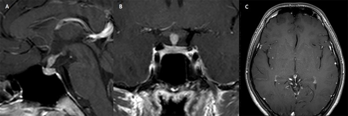
We recruited 158 patients with thickened pituitary stalk lesions visible on MRI. Laboratory findings included hypopituitarism, diabetes insipidus (DI), and hyperprolactinemia. MR images were assessed for anterior-posterior thickness (mm), diffuse pituitary stalk thickening, cystic changes, a high T1 signal, and glandular or extrasellar involvement. A diagnostic model was developed using a recursive partitioning logistic regression analysis. The model was validated in an independent dataset comprising 63 patients, and its diagnostic performance was compared with that of the original radiological reports.

RESULTS

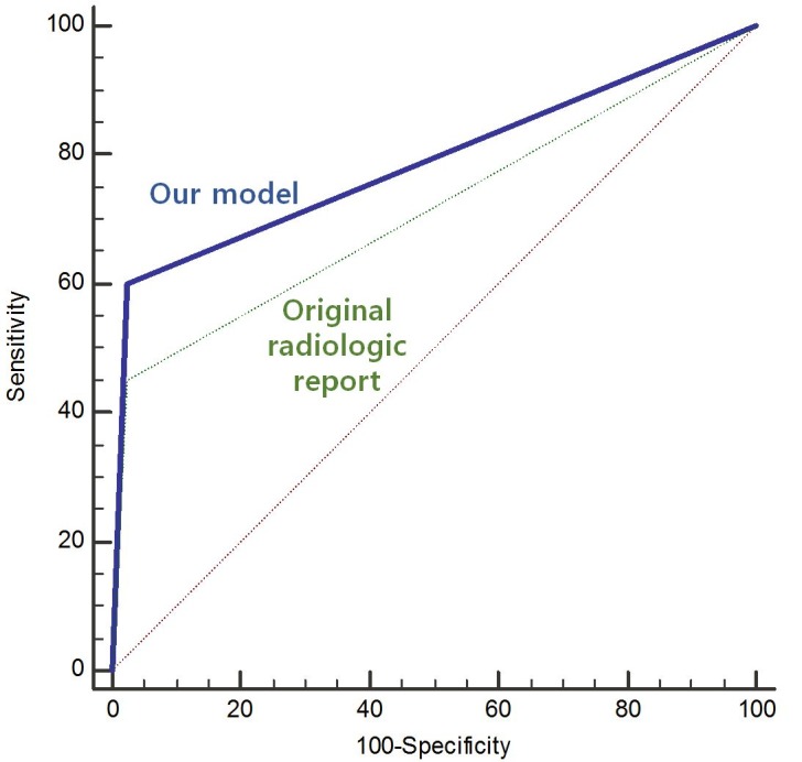
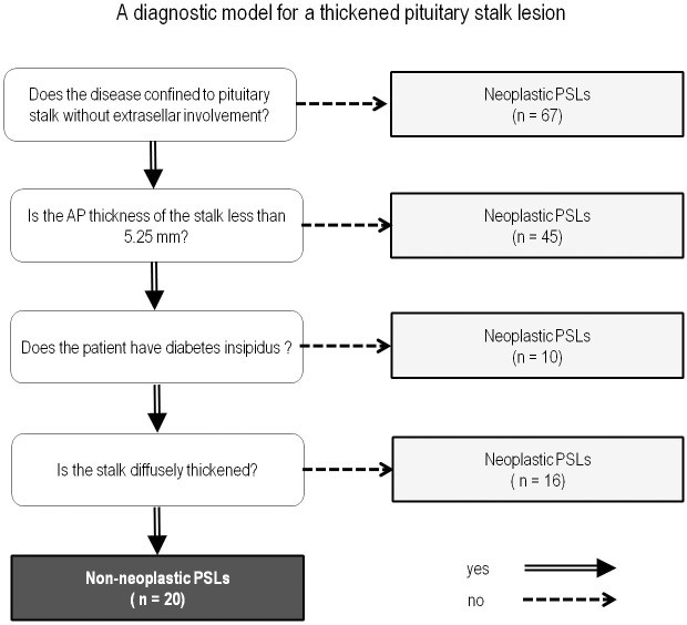
A univariate analysis found significant associations of DI (P = 0.006), absence of extrasellar involvement (P = 0.002), and lower stalk thickness (P = 0.031) with non-NPSLs. A diagnostic model was created using the following parameters (in order of priority): 1) lack of extrasellar involvement, 2) stalk thickness < 5.3 mm, and 3) presence of DI. The diagnostic performance (area under the curve; AUC) of this model in the independent set was 0.813, representing a significant improvement over the original radiological reports (AUC: 0.713, P = 0.029).

CONCLUSION

The joint diagnostic approach based on clinical and imaging-based factors robustly distinguished non-NPSLs from NPSLs. This approach could guide treatment strategies and prevent unnecessary surgery in patients with non-NPSL.

摘要

目的

区分非肿瘤性垂体柄病变(non-NPSLs)与肿瘤性垂体柄病变(NPSLs)是指导垂体柄增粗治疗的主要关注点。我们的研究旨在通过结合临床和磁共振成像(MRI)结果来区分non-NPSLs与NPSLs,以提供术前诊断帮助。

材料与方法

我们招募了158例MRI可见垂体柄增粗病变的患者。实验室检查结果包括垂体功能减退、尿崩症(DI)和高催乳素血症。对MR图像评估前后径厚度(mm)、垂体柄弥漫性增粗、囊性改变、T1高信号以及腺体内或鞍外受累情况。使用递归划分逻辑回归分析建立诊断模型。该模型在包含63例患者的独立数据集中进行验证,并将其诊断性能与原始放射学报告的诊断性能进行比较。

结果

单因素分析发现DI(P = 0.006)、无鞍外受累(P = 0.002)和较低的垂体柄厚度(P = 0.031)与non-NPSLs显著相关。使用以下参数(按优先级顺序)创建诊断模型:1)无鞍外受累,2)垂体柄厚度<5.3 mm,3)存在DI。该模型在独立数据集中的诊断性能(曲线下面积;AUC)为0.813,相较于原始放射学报告(AUC:0.713,P = 0.029)有显著改善。

结论

基于临床和影像学因素的联合诊断方法能有力地区分non-NPSLs与NPSLs。这种方法可指导治疗策略,并防止non-NPSL患者进行不必要的手术。

https://cdn.ncbi.nlm.nih.gov/pmc/blobs/20af/5687750/fbf46943f219/pone.0187989.g005.jpg
https://cdn.ncbi.nlm.nih.gov/pmc/blobs/20af/5687750/9ade224dfa41/pone.0187989.g001.jpg
https://cdn.ncbi.nlm.nih.gov/pmc/blobs/20af/5687750/4b243651e177/pone.0187989.g002.jpg
https://cdn.ncbi.nlm.nih.gov/pmc/blobs/20af/5687750/4455224de701/pone.0187989.g004.jpg
https://cdn.ncbi.nlm.nih.gov/pmc/blobs/20af/5687750/fbf46943f219/pone.0187989.g005.jpg

相似文献

1
Joint approach based on clinical and imaging features to distinguish non-neoplastic from neoplastic pituitary stalk lesions.基于临床和影像学特征的联合方法,用于区分非肿瘤性与肿瘤性垂体柄病变。
PLoS One. 2017 Nov 15;12(11):e0187989. doi: 10.1371/journal.pone.0187989. eCollection 2017.
2
Pituitary stalk lesions: the Mayo Clinic experience.垂体柄病变:梅奥诊所的经验。
J Clin Endocrinol Metab. 2013 May;98(5):1812-8. doi: 10.1210/jc.2012-4171. Epub 2013 Mar 26.
3
Central diabetes insipidus and pituitary stalk thickening in adults: distinction of neoplastic from non-neoplastic lesions.成人中枢性尿崩症与垂体柄增粗:鉴别肿瘤性与非肿瘤性病变。
Eur J Endocrinol. 2020 Jul 1;181(3):95-105. doi: 10.1530/EJE-20-0058.
4
Prediction of postoperative diabetes insipidus using morphological hyperintensity patterns in the pituitary stalk on magnetic resonance imaging after transsphenoidal surgery for sellar tumors.经蝶窦手术治疗鞍区肿瘤后,基于垂体柄磁共振成像形态学高信号模式预测术后尿崩症。
Pituitary. 2016 Dec;19(6):552-559. doi: 10.1007/s11102-016-0739-9.
5
Transsphenoidal Posterior Pituitary Lobe Biopsy in Patients with Neurohypophysial Lesions.患有神经垂体病变患者的经蝶窦垂体后叶活检
World Neurosurg. 2017 Mar;99:543-547. doi: 10.1016/j.wneu.2016.12.080. Epub 2016 Dec 27.
6
Pituitary stalk thickening: the role of an innovative MRI imaging analysis which may assist in determining clinical management.垂体柄增粗:一种创新的MRI成像分析的作用,其可能有助于确定临床管理。
Eur J Endocrinol. 2016 Oct;175(4):255-63. doi: 10.1530/EJE-16-0455. Epub 2016 Jul 14.
7
PITUITARY STALK THICKENING IN A LARGE COHORT: TOWARD MORE ACCURATE PREDICTORS OF PITUITARY DYSFUNCTION AND ETIOLOGY.大样本垂体柄增粗:更准确预测垂体功能障碍及病因。
Endocr Pract. 2019 Jun;25(6):534-544. doi: 10.4158/EP-2018-0550. Epub 2019 Mar 13.
8
Clinical and radiological features of pituitary stalk lesions in children and adolescents.儿童和青少年垂体柄病变的临床及影像学特征
Ann Pediatr Endocrinol Metab. 2014 Dec;19(4):202-7. doi: 10.6065/apem.2014.19.4.202. Epub 2014 Dec 31.
9
[Panhypopituitarism from pituitary metastases of breast cancer].[乳腺癌垂体转移导致的全垂体功能减退症]
Recenti Prog Med. 2007 Feb;98(2):87-9.
10
Thickened pituitary stalk on magnetic resonance imaging in children with central diabetes insipidus.中枢性尿崩症患儿磁共振成像显示垂体柄增粗。
Horm Res. 2000;53 Suppl 3:61-4. doi: 10.1159/000023536.

引用本文的文献

1
Complications of Endoscopic Skull Base Surgery for Sellar and Parasellar Tumors in Pediatric Population; Neurosurgical Perspectives.小儿鞍区及鞍旁肿瘤内镜颅底手术的并发症;神经外科观点
Front Oncol. 2022 May 27;12:769576. doi: 10.3389/fonc.2022.769576. eCollection 2022.
2
Safety of endoscopic endonasal biopsy for the pituitary stalk-hypothalamic lesions.经鼻内镜下对垂体柄-下丘脑病变进行活检的安全性。
Pituitary. 2022 Feb;25(1):143-151. doi: 10.1007/s11102-021-01181-0. Epub 2021 Sep 1.

本文引用的文献

1
Differentiation between Cystic Pituitary Adenomas and Rathke Cleft Cysts: A Diagnostic Model Using MRI.囊性垂体腺瘤与拉克氏裂囊肿的鉴别:一种使用MRI的诊断模型
AJNR Am J Neuroradiol. 2015 Oct;36(10):1866-73. doi: 10.3174/ajnr.A4387. Epub 2015 Aug 6.
2
Lymphocytic infundibulo-neurohypophysitis: a clinical overview.淋巴细胞性漏斗神经垂体炎:临床概述
Endocrine. 2015 Dec;50(3):531-6. doi: 10.1007/s12020-015-0707-6. Epub 2015 Jul 29.
3
Clinical and radiological features of pituitary stalk lesions in children and adolescents.
儿童和青少年垂体柄病变的临床及影像学特征
Ann Pediatr Endocrinol Metab. 2014 Dec;19(4):202-7. doi: 10.6065/apem.2014.19.4.202. Epub 2014 Dec 31.
4
Pituitary-Targeted Dynamic Contrast-Enhanced Multisection CT for Detecting MR Imaging-Occult Functional Pituitary Microadenoma.垂体靶向动态对比增强多层面CT用于检测磁共振成像隐匿性功能性垂体微腺瘤
AJNR Am J Neuroradiol. 2015 May;36(5):904-8. doi: 10.3174/ajnr.A4220. Epub 2015 Jan 15.
5
Pituitary stalk lesions: the Mayo Clinic experience.垂体柄病变:梅奥诊所的经验。
J Clin Endocrinol Metab. 2013 May;98(5):1812-8. doi: 10.1210/jc.2012-4171. Epub 2013 Mar 26.
6
Hypothalamo-pituitary sarcoidosis: a multicenter study of 24 patients.下丘脑-垂体结节病:24 例患者的多中心研究。
QJM. 2012 Oct;105(10):981-95. doi: 10.1093/qjmed/hcs121. Epub 2012 Jun 30.
7
Diagnosis and treatment of hyperprolactinemia: an Endocrine Society clinical practice guideline.高泌乳素血症的诊断和治疗:内分泌学会临床实践指南。
J Clin Endocrinol Metab. 2011 Feb;96(2):273-88. doi: 10.1210/jc.2010-1692.
8
Surgical treatment of pituitary tumours.垂体瘤的外科治疗。
Best Pract Res Clin Endocrinol Metab. 2009 Oct;23(5):677-92. doi: 10.1016/j.beem.2009.05.002.
9
Normal pituitary stalk: high-resolution MR imaging at 3T.正常垂体柄:3T 高分辨率磁共振成像。
AJNR Am J Neuroradiol. 2010 Feb;31(2):355-9. doi: 10.3174/ajnr.A1836. Epub 2009 Oct 1.
10
A radiologic score to distinguish autoimmune hypophysitis from nonsecreting pituitary adenoma preoperatively.术前鉴别自身免疫性垂体炎与无分泌功能垂体腺瘤的影像学评分。
AJNR Am J Neuroradiol. 2009 Oct;30(9):1766-72. doi: 10.3174/ajnr.A1714. Epub 2009 Jul 23.