Department of Radiology, Washington University School of Medicine, Saint Louis, Missouri, United States of America.
Department of Neurology, Washington University School of Medicine, Saint Louis, Missouri, United States of America.
PLoS One. 2017 Nov 17;12(11):e0188122. doi: 10.1371/journal.pone.0188122. eCollection 2017.
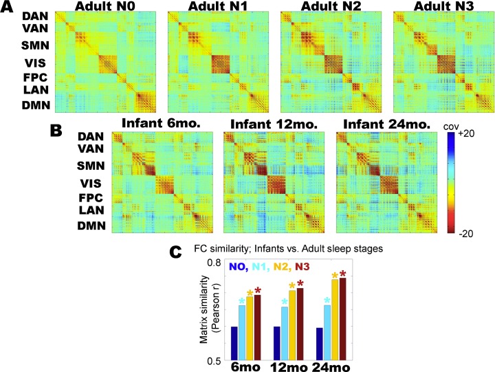
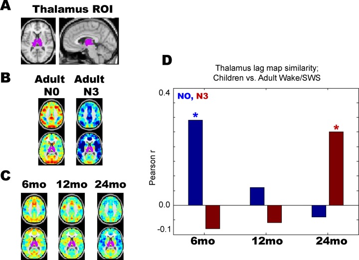
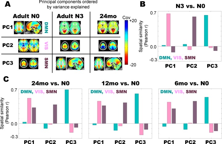
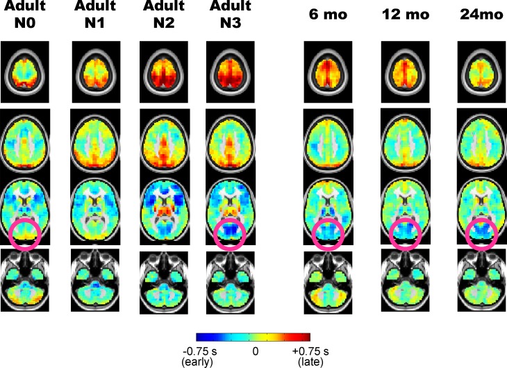
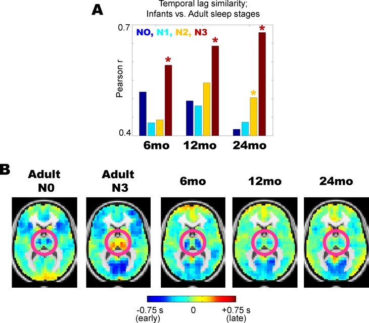
Resting state functional magnetic resonance imaging (rs-fMRI) in infants enables important studies of functional brain organization early in human development. However, rs-fMRI in infants has universally been obtained during sleep to reduce participant motion artifact, raising the question of whether differences in functional organization between awake adults and sleeping infants that are commonly attributed to development may instead derive, at least in part, from sleep. This question is especially important as rs-fMRI differences in adult wake vs. sleep are well documented. To investigate this question, we compared functional connectivity and BOLD signal propagation patterns in 6, 12, and 24 month old sleeping infants with patterns in adult wakefulness and non-REM sleep. We find that important functional connectivity features seen during infant sleep closely resemble those seen during adult sleep, including reduced default mode network functional connectivity. However, we also find differences between infant and adult sleep, especially in thalamic BOLD signal propagation patterns. These findings highlight the importance of considering sleep state when drawing developmental inferences in infant rs-fMRI.
静息态功能磁共振成像 (rs-fMRI) 在婴儿中得到了广泛应用,可用于在人类发育早期研究大脑功能组织。然而,为了减少被试者运动伪影,婴儿的 rs-fMRI 普遍是在睡眠中获得的,这就提出了一个问题,即通常归因于发育的清醒成年人和睡眠中婴儿之间的功能组织差异,是否至少部分源自睡眠。这个问题尤为重要,因为成人清醒和睡眠状态下 rs-fMRI 的差异已有充分的记录。为了研究这个问题,我们比较了 6、12 和 24 个月大的睡眠中婴儿与成人清醒和非快速眼动 (NREM) 睡眠状态下的功能连接和 BOLD 信号传播模式。我们发现,在婴儿睡眠中观察到的重要功能连接特征与在成人睡眠中观察到的特征非常相似,包括默认模式网络功能连接的减少。然而,我们也发现了婴儿和成人睡眠之间的差异,特别是在丘脑 BOLD 信号传播模式上。这些发现强调了在对婴儿 rs-fMRI 进行发育推断时考虑睡眠状态的重要性。