Department of Nuclear Medicine, Institut de Diagnòstic per la Imatge (IDI), Hospital General Universitari Vall d'Hebrón, Universitat Autònoma de Barcelona, Barcelona, Spain.
Department of Nuclear Medicine, Gammagrafía Corachan, Barcelona, Spain.
J Alzheimers Dis. 2018;61(1):321-332. doi: 10.3233/JAD-170693.
Recently, modifications of Aβ1-42 levels in CSF and plasma associated with improvement in memory and language functions have been observed in patients with mild-moderate Alzheimer's disease (AD) treated with plasma exchange (PE) with albumin replacement.
To detect structural and functional brain changes in PE-treated AD patients as part of a Phase II clinical trial.
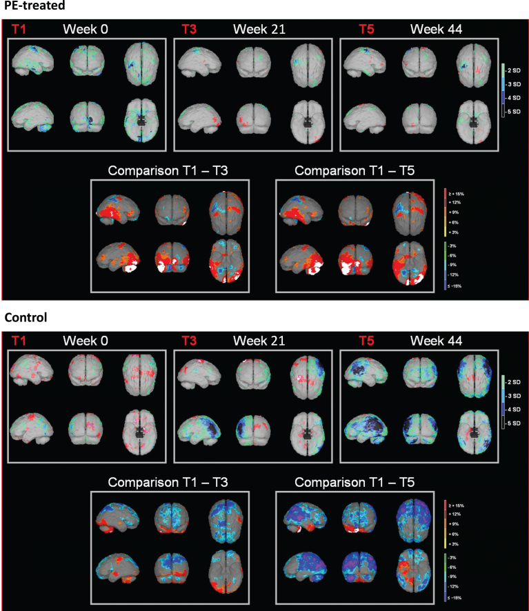
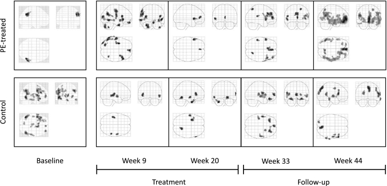
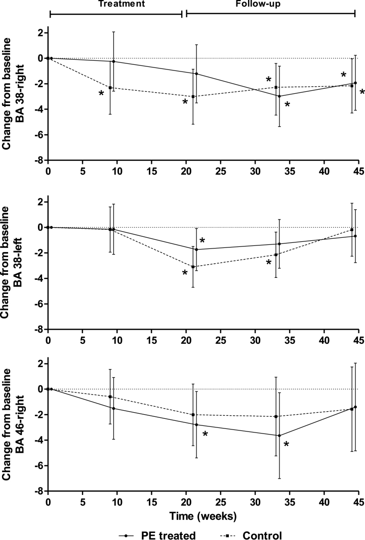
Patients received between 3 and 18 PE with albumin (Albutein® 5%, Grifols) or sham-PE (controls) for 21 weeks (divided in one intensive and two maintenance periods) followed by 6-month follow-up. Brain perfusion assessed by SPECT scans using an automated software (NeuroGam®) and brain structural changes assessed by MRI were performed at weeks 0 (baseline), 21, and 44 (with additional SPECT at weeks 9 and 33). Statistical parametric mapping (voxel-based analysis, SPM) and Z-scores calculations were applied to investigate changes to baseline.
42 patients were recruited (39 evaluable; 37 analyzed: 18 PE-treated; 19 controls). There was a trend toward decreasing hippocampi and total intracranial volume for both patient groups during the study (p < 0.05). After six months, PE-treated patients had less cerebral perfusion loss than controls in frontal, temporal, and parietal areas, and perfusion stabilization in Brodmann area BA38-R during the PE-treatment period (p < 0.05). SPM analysis showed stabilization or absence of progression of perfusion loss in PE-treated patients until week 21, not observed in controls.
Mild-moderate AD patients showed decreased brain volume and impairment of brain perfusion as expected for the progression of the disease. PE-treatment with albumin replacement favored the stabilization of perfusion.
最近,在接受白蛋白置换的血浆置换 (PE) 治疗的轻度至中度阿尔茨海默病 (AD) 患者中,观察到 CSF 和血浆中 Aβ1-42 水平的改变与记忆和语言功能的改善有关。
检测 PE 治疗 AD 患者的大脑结构和功能变化,作为 II 期临床试验的一部分。
患者接受了 3 到 18 次白蛋白 (Albutein® 5%,Grifols) 或假 PE (对照组) 的 PE 治疗,共 21 周(分为一个强化期和两个维持期),然后进行 6 个月的随访。使用自动软件 (NeuroGam®) 进行 SPECT 扫描评估脑灌注,使用 MRI 评估脑结构变化,在第 0 周(基线)、21 周和 44 周(在第 9 周和 33 周进行额外的 SPECT)进行评估。应用统计参数映射 (voxel-based analysis, SPM) 和 Z 分数计算来研究对基线的变化。
共招募了 42 名患者(39 名可评估;37 名进行了分析:18 名接受 PE 治疗;19 名对照组)。在研究期间,两组患者的海马体和总颅内体积都有下降的趋势(p<0.05)。六个月后,与对照组相比,PE 治疗组患者在额叶、颞叶和顶叶区域的脑灌注损失较少,并且在 PE 治疗期间 Brodmann 区域 BA38-R 的灌注稳定(p<0.05)。SPM 分析显示,PE 治疗组患者的灌注损失稳定或没有进展,而对照组则没有。
轻度至中度 AD 患者的脑体积减少,脑灌注受损,符合疾病进展的预期。用白蛋白替代物进行 PE 治疗有利于灌注的稳定。