Bittner-Eddy Peter D, Fischer Lori A, Tu Andy A, Allman Daniel A, Costalonga Massimo
Division of Periodontology, Department of Developmental and Surgical Sciences, School of Dentistry, University of Minnesota, Minneapolis, MN, United States.
Front Immunol. 2017 Oct 30;8:1398. doi: 10.3389/fimmu.2017.01398. eCollection 2017.
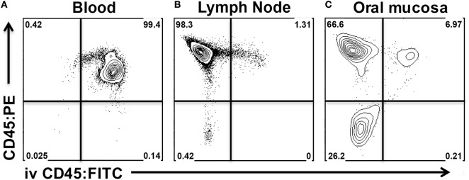
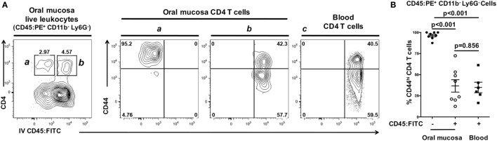
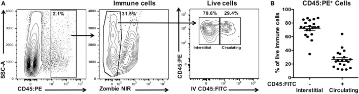
Periodontitis is a chronic inflammatory response to a microbial biofilm that destroys bone and soft tissues supporting the teeth. Murine models of periodontitis based on () colonization have shown that extravasation of leukocytes into oral tissue is critical to driving alveolar bone destruction. Identifying interstitial leukocytes is key to understanding the immunopathogenesis of periodontitis. Here, we describe a robust flow cytometry assay based on intravenous FITC-conjugated anti-mouse CD45 mAb that distinguishes interstitial leukocytes in the oral mucosa of mice from those circulating within the vasculature or in post-dissection contaminating blood. Unaccounted circulating leukocytes skewed the relative frequency of B cells and granulocytes and inflated the numbers of all leukocyte cell types. We also describe a dissection technique that avoids contamination of oral mucosal tissues with nasal-associated lymphoid tissues (NALT), a B cell rich organ that can inflate leukocyte numbers at least 10-fold and skew the assessment of interstitial CD4 T cell phenotypes. Unlike circulating CD4 T cells, interstitial CD4 T cells were almost exclusively antigen-experienced cells (CD44). We report for the first time the presence of antigen-experienced -specific CD4 T cells in NALT following oral feeding of mice with . This new combined flow cytometry and dissection approach allows identification of leukocytes infiltrating the connective tissues of the murine oral mucosa and avoids confounding analyses of leukocytes not recruited to inflamed oral mucosal tissues in disease conditions like periodontitis, candidiasis, or sialadenitis.
牙周炎是对微生物生物膜的慢性炎症反应,该生物膜会破坏支持牙齿的骨骼和软组织。基于()定植的牙周炎小鼠模型表明,白细胞渗入口腔组织对于驱动牙槽骨破坏至关重要。识别间质白细胞是理解牙周炎免疫发病机制的关键。在此,我们描述了一种基于静脉注射异硫氰酸荧光素(FITC)偶联的抗小鼠CD45单克隆抗体的强大流式细胞术检测方法,该方法可区分小鼠口腔黏膜中的间质白细胞与血管内循环的白细胞或解剖后污染血液中的白细胞。未计入的循环白细胞会使B细胞和粒细胞的相对频率发生偏差,并使所有白细胞类型的数量膨胀。我们还描述了一种解剖技术,可避免口腔黏膜组织被鼻相关淋巴组织(NALT)污染,NALT是一个富含B细胞的器官,它可使白细胞数量至少增加10倍,并扭曲间质CD4 T细胞表型的评估。与循环CD4 T细胞不同,间质CD4 T细胞几乎完全是经历过抗原刺激的细胞(CD44)。我们首次报告了在用()口服喂养小鼠后,NALT中存在经历过抗原刺激的特异性CD4 T细胞。这种新的流式细胞术与解剖相结合的方法能够识别渗入小鼠口腔黏膜结缔组织的白细胞,并避免在牙周炎、念珠菌病或涎腺炎等疾病状态下对未募集到发炎口腔黏膜组织中的白细胞进行混淆分析。