Cha Yoon Jin, Shim Hyo Sup
Department of Pathology, Severance Hospital, Yonsei University College of Medicine, Seoul, Korea.
Oncotarget. 2017 Sep 15;8(52):89465-89474. doi: 10.18632/oncotarget.20948. eCollection 2017 Oct 27.
Inflammatory myofibroblastic tumors (IMTs) are rare mesenchymal neoplasms that are composed of myofibroblastic cells accompanied by inflammatory infiltrate. We investigated the immune profiles of IMTs, including PD-L1 expression and proportion of CD8+ tumor-infiltrating lymphocytes (TILs), as well as its clinicopathological characteristics according to gene rearrangementstatus.
Twenty-eight IMTs from 25 patients were retrieved from our pathology files (2005-2015), and their clinicopathological parameters and outcomes were analyzed. Immunohistochemistry (IHC) was performed using whole-tissue sections to detect PD-L1 and CD8 expression, and fluorescent in situ hybridization (FISH) analysis and IHC were performed using tissue microarrays to identify rearrangements in the , and genes.
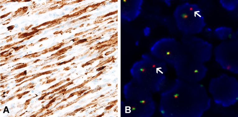
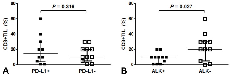
rearrangement was observed in 11 cases (44.0%), and all cases exhibited diffuse cytoplasmic expression during IHC. or rearrangement was not detected using IHC or FISH. IMTs harboring rearrangement (-positive) were located in the lungs ( = 7), genitourinary tract ( = 2), and mesentery ( = 1). The mean patient age was 33.2 years for -positive IMTs and 53.1 years for -negative IMTs. All patients with -positive IMTs survived without recurrence or metastasis. IMTs with metastasis and/or recurrence were -negative and exhibited elevated PD-L1 expression (positive tumor cells: 70.0% . 21.3%, = 0.023; H-score: 107.5 . 26.3, = 0.005). In addition, -negative IMTs had a more CD8+ TILs, compared to -positive IMTs (23.3% . 8.9%, = 0.027).
-positive IMTs are characterized by younger age, well-defined margins, frequent involvement of the lung, and fewer CD8+ TILs. Greater PD-L1 expression was observed in IMTs with tumor necrosis and metastasis/recurrence, which were also negative for rearrangement. These results suggest that immune checkpoint inhibitors may be a novel option for treating patients with advanced IMT.
炎性肌纤维母细胞瘤(IMTs)是一种罕见的间叶性肿瘤,由肌纤维母细胞组成,并伴有炎性浸润。我们研究了IMTs的免疫特征,包括程序性死亡受体配体1(PD-L1)表达和CD8 +肿瘤浸润淋巴细胞(TILs)比例,以及根据基因重排状态的临床病理特征。
从我们的病理档案(2005 - 2015年)中检索出25例患者的28个IMTs,并分析其临床病理参数和预后。使用全组织切片进行免疫组织化学(IHC)检测PD-L1和CD8表达,使用组织微阵列进行荧光原位杂交(FISH)分析和IHC,以鉴定ALK、ROS1和RET基因的重排。
11例(44.0%)观察到ALK重排,所有病例在IHC期间均表现为弥漫性细胞质ALK表达。使用IHC或FISH未检测到ROS1或RET重排。携带ALK重排(ALK阳性)的IMTs位于肺部(n = 7)、泌尿生殖道(n = 2)和肠系膜(n = 1)。ALK阳性IMTs患者的平均年龄为33.2岁,ALK阴性IMTs患者的平均年龄为53.1岁。所有ALK阳性IMTs患者均存活,无复发或转移。发生转移和/或复发的IMTs为ALK阴性,且PD-L1表达升高(阳性肿瘤细胞:70.0%对21.3%,P = 0.023;H评分:107.5对26.3,P = 0.005)。此外,与ALK阳性IMTs相比,ALK阴性IMTs的CD8 + TILs更多(23.3%对8.9%,P = 0.027)。
ALK阳性IMTs的特征是年龄较小、边界清晰、肺部受累频繁且CD8 + TILs较少。在伴有肿瘤坏死和转移/复发的IMTs中观察到更高的PD-L1表达,这些IMTs的ALK重排也为阴性。这些结果表明,免疫检查点抑制剂可能是治疗晚期IMTs患者的一种新选择。