Marcq Elly, Waele Jorrit De, Audenaerde Jonas Van, Lion Eva, Santermans Eva, Hens Niel, Pauwels Patrick, van Meerbeeck Jan P, Smits Evelien L J
Center for Oncological Research, University of Antwerp, Antwerp, Belgium.
Laboratory of Experimental Hematology, University of Antwerp, Antwerp, Belgium.
Oncotarget. 2017 Sep 21;8(52):89722-89735. doi: 10.18632/oncotarget.21113. eCollection 2017 Oct 27.
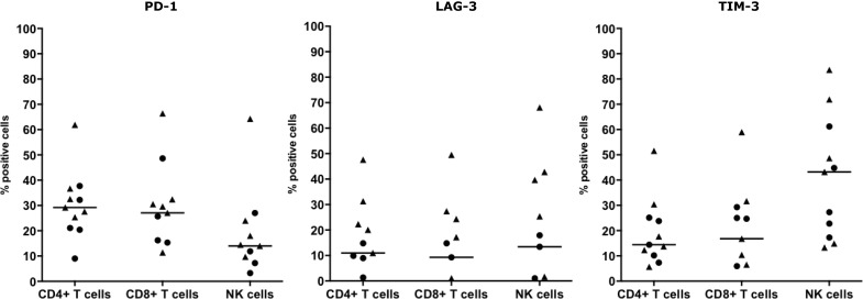
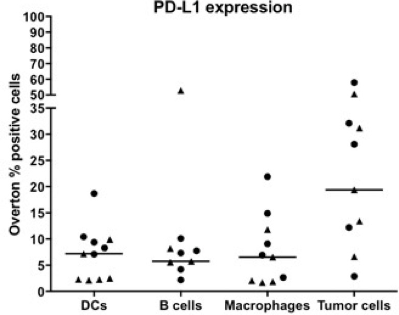
Malignant pleural mesothelioma (MPM) is an aggressive cancer with an increasing incidence, poor prognosis and limited effective treatment options. Hence, new treatment strategies are warranted which include immune checkpoint blockade approaches with encouraging preliminary data. Research on the immunological aspects of the easily accessible mesothelioma microenvironment could identify prognostic and/or predictive biomarkers and provide useful insights for developing effective immunotherapy. In this context, we investigated the immune cell composition of effusions (pleural and ascites fluids) from 11 different chemotherapy-treated MPM patients. We used multicolor flow cytometry to describe different subsets of immune cells and their expression of immune checkpoint molecules TIM-3, LAG-3, PD-1 and PD-L1. We demonstrate a patient-dependent inter- and intraspecific variation comparing pleural and ascites fluids in immune cell composition and immune checkpoint expression. We found CD4 and CD8 T cells, B cells, macrophages, natural killer cells, dendritic cells and tumor cells in the fluids. To the best of our knowledge, we are the first to report TIM-3 and LAG-3 expression and we confirm PD-1 and PD-L1 expression on different MPM effusion-resident immune cells. Moreover, we identified two MPM effusion-related factors with clinical value: CD4+ T cells were significantly correlated with better response to chemotherapy, while the percentage of PD-L1 podoplanin (PDPN) tumor cells is a significant prognostic factor for worse outcome. Our data provide a basis for more elaborate research on MPM effusion material in the context of treatment follow-up and prognostic biomarkers and the development of immune checkpoint-targeted immunotherapy.
恶性胸膜间皮瘤(MPM)是一种侵袭性癌症,发病率不断上升,预后较差,有效治疗选择有限。因此,需要新的治疗策略,其中包括免疫检查点阻断方法,其初步数据令人鼓舞。对易于获取的间皮瘤微环境的免疫学方面进行研究,可能会识别出预后和/或预测生物标志物,并为开发有效的免疫疗法提供有用的见解。在此背景下,我们调查了11名接受不同化疗的MPM患者的胸腔积液(胸水和腹水)中的免疫细胞组成。我们使用多色流式细胞术来描述免疫细胞的不同亚群及其免疫检查点分子TIM-3、LAG-3、PD-1和PD-L1的表达。我们证明,在免疫细胞组成和免疫检查点表达方面,胸水和腹水之间存在患者依赖的种间和种内差异。我们在这些液体中发现了CD4和CD8 T细胞、B细胞、巨噬细胞、自然杀伤细胞、树突状细胞和肿瘤细胞。据我们所知,我们是第一个报告TIM-3和LAG-3表达的,并证实了不同MPM积液驻留免疫细胞上PD-1和PD-L1的表达。此外,我们确定了两个具有临床价值的MPM积液相关因素:CD4+ T细胞与化疗更好的反应显著相关,而PD-L1足板蛋白(PDPN)肿瘤细胞的百分比是预后较差的一个重要预后因素。我们的数据为在治疗随访、预后生物标志物以及免疫检查点靶向免疫疗法开发的背景下,对MPM积液材料进行更深入研究提供了基础。