Kulikov Alexei N, Sosnovskii Sergei V, Berezin Roman D, Maltsev Dmitrii S, Oskanov Dzhambulat H, Gribanov Nikolai A
Department of Ophthalmology, Military Medical Academy, St Petersburg, Russia.
Clin Ophthalmol. 2017 Nov 14;11:1995-2002. doi: 10.2147/OPTH.S146019. eCollection 2017.
To study vitreoretinal interface (VRI) abnormalities in diabetic macular edema (DME) and the influence of these on the effectiveness of intravitreal anti-vascular endothelial growth factor (VEGF) therapy.
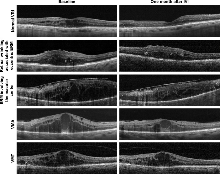
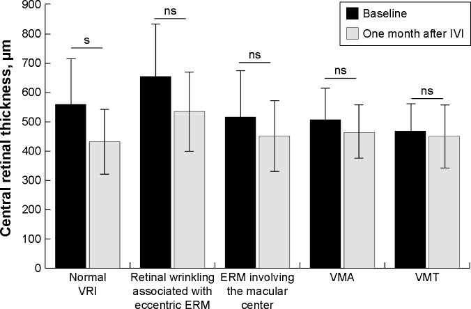
VRI status and central retinal thickness (CRT) were evaluated using line and 3D-reference scans obtained using spectral domain-optical coherence tomography RTVue-100 before and 1 month after intravitreal anti-VEGF injection (IVI). VRI status was categorized into five subgroups: normal VRI, retinal surface wrinkling associated with the eccentric epiretinal membrane (ERM), ERM involving the macular center, vitreomacular adhesion (VMA), and vitreomacular traction (VMT).
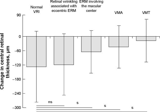
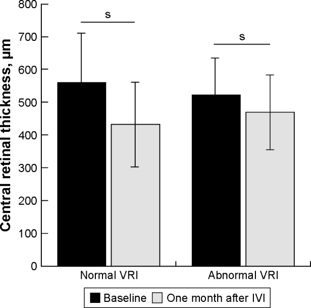
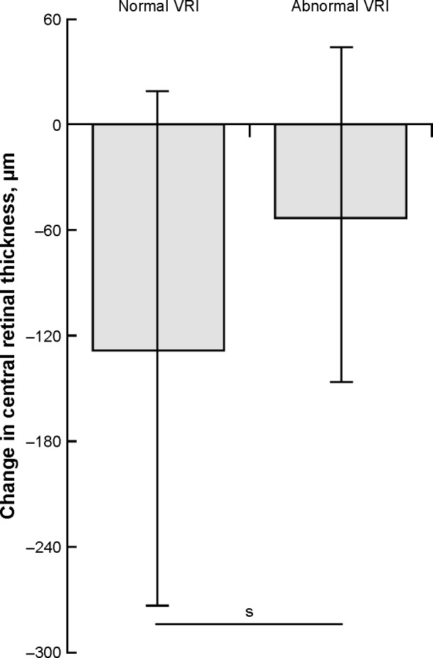
A total of 105 eyes of 89 patients were included in the study. One month after IVI, the mean change of CRT in normal VRI eyes and eyes with VRI abnormalities was -128.0±144.7 µm and -53.0±96.4 µm (<0.05), respectively. The mean change of CRT 1 month after IVI in each subgroup with VRI abnormalities, apart from the subgroup with retinal wrinkling associated with eccentric ERM, was statistically significantly lower compared to the eyes with normal VRI (<0.05).
VRI abnormalities significantly reduce the effectiveness of intravitreal anti-VEGF therapy in eyes with DME. Eyes with noticeable changes of VRI, including ERM involving the macular center, VMA, and VMT have a poorer response to anti-VEGF therapy compared to eyes with normal VRI or eccentric ERM.
研究糖尿病性黄斑水肿(DME)患者的玻璃体视网膜界面(VRI)异常情况及其对玻璃体内抗血管内皮生长因子(VEGF)治疗效果的影响。
在玻璃体内注射抗VEGF(IVI)前及注射后1个月,使用光谱域光学相干断层扫描RTVue - 100获得的线性和三维参考扫描评估VRI状态和中心视网膜厚度(CRT)。VRI状态分为五个亚组:正常VRI、与偏心性视网膜前膜(ERM)相关的视网膜表面皱襞、累及黄斑中心的ERM、玻璃体黄斑粘连(VMA)和玻璃体黄斑牵引(VMT)。
本研究共纳入89例患者的105只眼。IVI后1个月,正常VRI眼和VRI异常眼的CRT平均变化分别为 - 128.0±144.7 µm和 - 53.0±96.4 µm(P<0.05)。除与偏心性ERM相关的视网膜皱襞亚组外,各VRI异常亚组IVI后1个月的CRT平均变化与正常VRI眼相比差异有统计学意义(P<0.05)。
VRI异常显著降低了DME患者玻璃体内抗VEGF治疗的效果。与正常VRI或偏心性ERM的眼相比,VRI有明显变化的眼,包括累及黄斑中心的ERM、VMA和VMT,对抗VEGF治疗的反应较差。