Computational, Cognitive and Clinical Neuroimaging Laboratory, Imperial College London, Division of Brain Sciences, Hammersmith Hospital, London, UK.
Department of Bioengineering, Imperial College London, London, UK.
Brain. 2018 Jan 1;141(1):148-164. doi: 10.1093/brain/awx309.
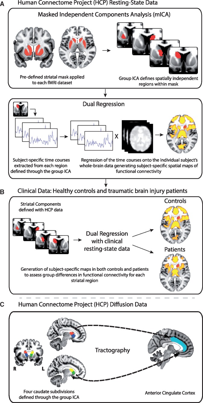
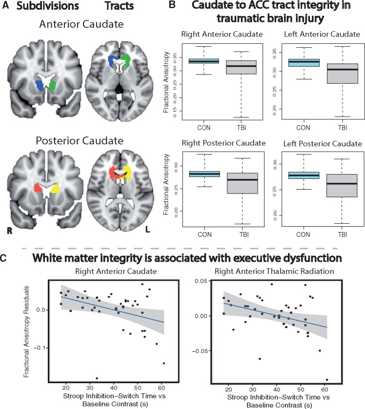
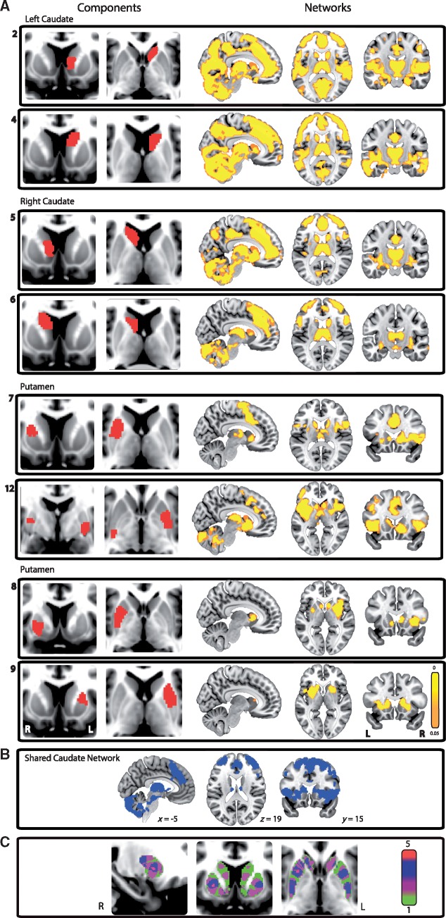
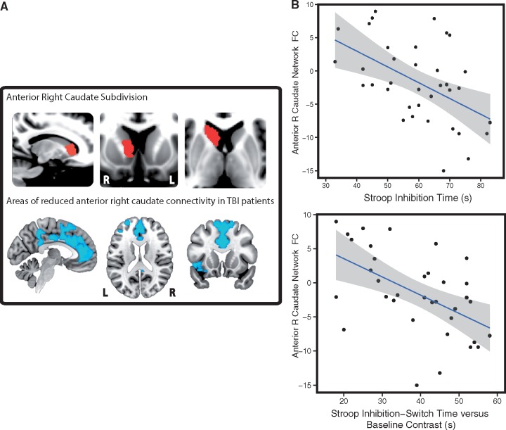
Traumatic brain injury often produces executive dysfunction. This characteristic cognitive impairment often causes long-term problems with behaviour and personality. Frontal lobe injuries are associated with executive dysfunction, but it is unclear how these injuries relate to corticostriatal interactions that are known to play an important role in behavioural control. We hypothesized that executive dysfunction after traumatic brain injury would be associated with abnormal corticostriatal interactions, a question that has not previously been investigated. We used structural and functional MRI measures of connectivity to investigate this. Corticostriatal functional connectivity in healthy individuals was initially defined using a data-driven approach. A constrained independent component analysis approach was applied in 100 healthy adult dataset from the Human Connectome Project. Diffusion tractography was also performed to generate white matter tracts. The output of this analysis was used to compare corticostriatal functional connectivity and structural integrity between groups of 42 patients with traumatic brain injury and 21 age-matched controls. Subdivisions of the caudate and putamen had distinct patterns of functional connectivity. Traumatic brain injury patients showed disruption to functional connectivity between the caudate and a distributed set of cortical regions, including the anterior cingulate cortex. Cognitive impairments in the patients were mainly seen in processing speed and executive function, as well as increased levels of apathy and fatigue. Abnormalities of caudate functional connectivity correlated with these cognitive impairments, with reductions in right caudate connectivity associated with increased executive dysfunction, information processing speed and memory impairment. Structural connectivity, measured using diffusion tensor imaging between the caudate and anterior cingulate cortex was impaired and this also correlated with measures of executive dysfunction. We show for the first time that altered subcortical connectivity is associated with large-scale network disruption in traumatic brain injury and that this disruption is related to the cognitive impairments seen in these patients.
创伤性脑损伤常导致执行功能障碍。这种特征性认知障碍常导致行为和人格的长期问题。额叶损伤与执行功能障碍有关,但尚不清楚这些损伤与皮质纹状体相互作用有何关系,皮质纹状体相互作用已知在行为控制中起着重要作用。我们假设创伤性脑损伤后的执行功能障碍与皮质纹状体相互作用异常有关,这是一个以前没有研究过的问题。我们使用结构和功能 MRI 连接度测量来对此进行研究。首先使用数据驱动的方法确定健康个体的皮质纹状体功能连接。在来自人类连接组计划的 100 个健康成人数据集上应用了受约束的独立成分分析方法。还进行了扩散轨迹追踪以生成白质束。该分析的结果用于比较创伤性脑损伤患者和 21 名年龄匹配对照组之间的皮质纹状体功能连接和结构完整性。尾状核和壳核的细分具有不同的功能连接模式。创伤性脑损伤患者表现出尾状核与包括前扣带皮层在内的皮质区域之间的功能连接中断。患者的认知障碍主要表现为处理速度和执行功能障碍,以及增加的淡漠和疲劳水平。尾状核功能连接的异常与这些认知障碍相关,右侧尾状核连接减少与执行功能障碍、信息处理速度和记忆障碍增加相关。使用弥散张量成像测量的尾状核和前扣带皮层之间的结构连接受损,这也与执行功能障碍的测量相关。我们首次表明,改变的皮质下连接与创伤性脑损伤中的大规模网络中断有关,这种中断与这些患者中出现的认知障碍有关。