Department of Pathology, Massachusetts General Hospital, Harvard Medical School, Boston, MA, USA.
Department of Radiology, Massachusetts General Hospital, Harvard Medical School, Boston, MA, USA.
Prostate Cancer Prostatic Dis. 2018 Sep;21(3):297-305. doi: 10.1038/s41391-017-0011-z. Epub 2017 Dec 5.
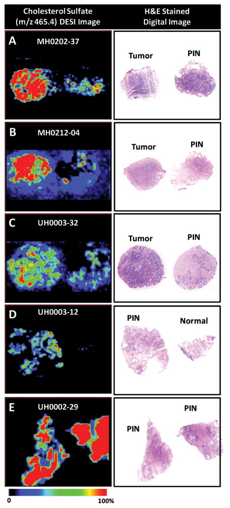
Prostate cancer (PCa), the most common cancer and second leading cause of cancer death in American men, presents the clinical challenge of distinguishing between indolent and aggressive tumors for proper treatment. PCa presents significant alterations in metabolic pathways that can potentially be measured using techniques like mass spectrometry (MS) or MS imaging (MSI) and used to characterize PCa aggressiveness. MS quantifies metabolomic, proteomic, and lipidomic profiles of biological systems that can be further visualized for their spatial distributions through MSI.
PubMed was queried for all publications relating to MS and MSI in human PCa from April 2007 to April 2017. With the goal of reviewing the utility of MSI in diagnosis and prognostication of human PCa, MSI articles that reported investigations of PCa-specific metabolites or metabolites indicating PCa aggressiveness were selected for inclusion. Articles were included that covered MS and MSI principles, limitations, and applications in PCa.
We identified nine key studies on MSI in intact human prostate tissue specimens that determined metabolites which could either differentiate between benign and malignant prostate tissue or indicate PCa aggressiveness. These MSI-detected biomarkers show promise in reliably identifying PCa and determining disease aggressiveness.
MSI represents an innovative technique with the ability to interrogate cancer biomarkers in relation to tissue pathologies and investigate tumor aggressiveness. We propose MSI as a powerful adjuvant histopathology imaging tool for prostate tissue evaluations, where clinical translation of this ex vivo technique could make possible the use of MSI for personalized medicine in diagnosis and prognosis of PCa. Moreover, the knowledge provided from this technique can majorly contribute to the understanding of molecular pathogenesis of PCa and other malignant diseases.
前列腺癌(PCa)是美国男性中最常见的癌症和第二大癌症死因,其临床挑战在于区分惰性和侵袭性肿瘤,以进行适当的治疗。PCa 在代谢途径中存在显著改变,这些改变可以通过质谱(MS)或 MS 成像(MSI)等技术进行测量,并用于表征 PCa 的侵袭性。MS 定量分析生物系统的代谢组学、蛋白质组学和脂质组学谱,通过 MSI 可以进一步可视化它们的空间分布。
在 2007 年 4 月至 2017 年 4 月期间,通过 PubMed 检索了所有与人类 PCa 中的 MS 和 MSI 相关的出版物。为了评估 MSI 在人类 PCa 的诊断和预后中的效用,选择了报道了 PCa 特异性代谢物或指示 PCa 侵袭性的代谢物的 MSI 文章进行纳入。纳入的文章涵盖了 MS 和 MSI 的原理、局限性及其在 PCa 中的应用。
我们确定了九项关于完整人类前列腺组织标本中 MSI 的关键研究,这些研究确定了可以区分良性和恶性前列腺组织或指示 PCa 侵袭性的代谢物。这些 MSI 检测到的生物标志物在可靠地识别 PCa 和确定疾病侵袭性方面显示出了潜力。
MSI 代表了一种创新技术,具有询问与组织病理学相关的癌症生物标志物并研究肿瘤侵袭性的能力。我们提出 MSI 作为一种强大的辅助组织病理学成像工具,用于前列腺组织评估,该技术的临床转化可能使 MSI 能够用于 PCa 的诊断和预后的个体化医学。此外,该技术提供的知识将极大地有助于理解 PCa 和其他恶性疾病的分子发病机制。