Department of Nuclear Medicine and Molecular Imaging, University Medical Center Groningen, University of Groningen, Groningen, The Netherlands.
J Neuroendocrinol. 2018 Feb;30(2). doi: 10.1111/jne.12565.
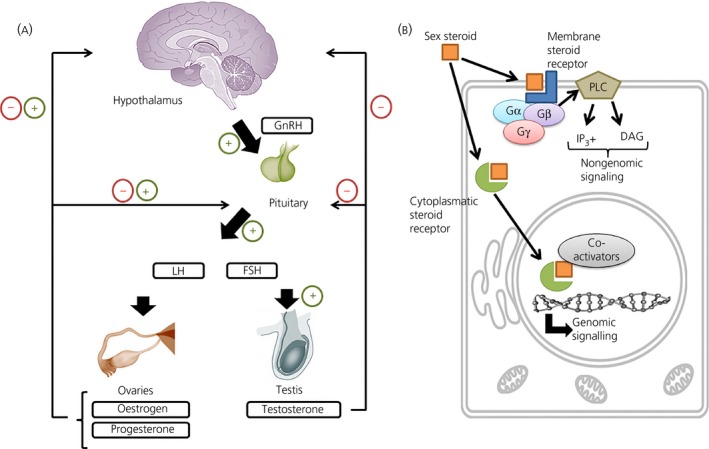
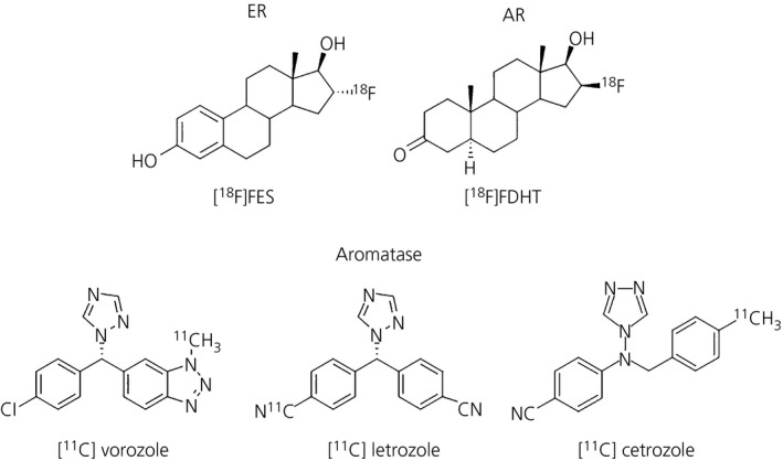
Sex steroid hormones are major regulators of sexual characteristic among species. These hormones, however, are also produced in the brain. Steroidal hormone-mediated signalling via the corresponding hormone receptors can influence brain function at the cellular level and thus affect behaviour and higher brain functions. Altered steroid hormone signalling has been associated with psychiatric disorders, such as anxiety and depression. Neurosteroids are also considered to have a neuroprotective effect in neurodegenerative diseases. So far, the role of steroid hormone receptors in physiological and pathological conditions has mainly been investigated post mortem on animal or human brain tissues. To study the dynamic interplay between sex steroids, their receptors, brain function and behaviour in psychiatric and neurological disorders in a longitudinal manner, however, non-invasive techniques are needed. Positron emission tomography (PET) is a non-invasive imaging tool that is used to quantitatively investigate a variety of physiological and biochemical parameters in vivo. PET uses radiotracers aimed at a specific target (eg, receptor, enzyme, transporter) to visualise the processes of interest. In this review, we discuss the current status of the use of PET imaging for studying sex steroid hormones in the brain. So far, PET has mainly been investigated as a tool to measure (changes in) sex hormone receptor expression in the brain, to measure a key enzyme in the steroid synthesis pathway (aromatase) and to evaluate the effects of hormonal treatment by imaging specific downstream processes in the brain. Although validated radiotracers for a number of targets are still warranted, PET can already be a useful technique for steroid hormone research and facilitate the translation of interesting findings in animal studies to clinical trials in patients.
性甾体激素是物种间性特征的主要调节者。然而,这些激素也在大脑中产生。类固醇激素通过相应的激素受体介导的信号可以在细胞水平上影响大脑功能,从而影响行为和更高的大脑功能。改变的类固醇激素信号与精神疾病有关,如焦虑和抑郁。神经甾体也被认为在神经退行性疾病中有神经保护作用。到目前为止,甾体激素受体在生理和病理条件下的作用主要是在动物或人类脑组织上进行死后研究。然而,为了研究性甾体、其受体、大脑功能和行为在精神和神经疾病中的动态相互作用,需要非侵入性技术。正电子发射断层扫描(PET)是一种非侵入性的成像工具,用于在体内定量研究各种生理和生化参数。PET 使用针对特定目标(例如受体、酶、转运体)的放射性示踪剂来可视化感兴趣的过程。在这篇综述中,我们讨论了使用 PET 成像研究大脑中性甾体激素的当前状况。到目前为止,PET 主要被用作测量大脑中性激素受体表达(变化)的工具,测量类固醇合成途径中的关键酶(芳香酶),并通过成像大脑中特定的下游过程来评估激素治疗的效果。尽管仍需要验证许多目标的放射性示踪剂,但 PET 已经可以成为研究类固醇激素的有用技术,并有助于将动物研究中的有趣发现转化为患者的临床试验。