Zhang Junjie, Cao Yanan, Gao Xiaowei, Zhu Maoen, Zhang Zhong, Yang Yue, Guo Qulian, Peng Yonggang, Wang E
1 159374 Department of Anesthesiology, Xiangya Hospital, Central South University, Changsha, Hunan, China.
2 Department of Anesthesiology, Shands Hospital, University of Florida, Gainesville, FL, USA.
Pulm Circ. 2018 Jan-Mar;8(1):2045893217744504. doi: 10.1177/2045893217744504. Epub 2017 Dec 18.
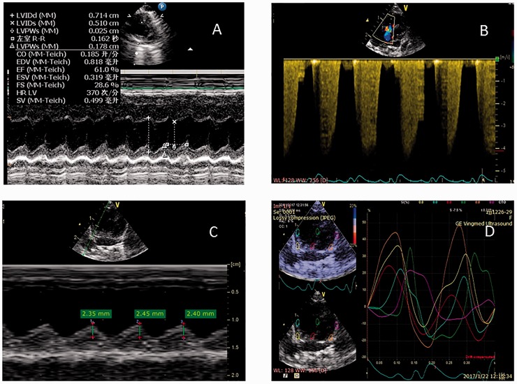
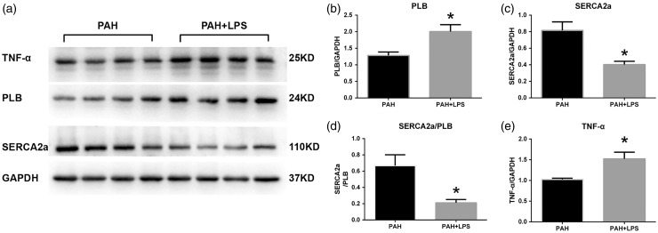
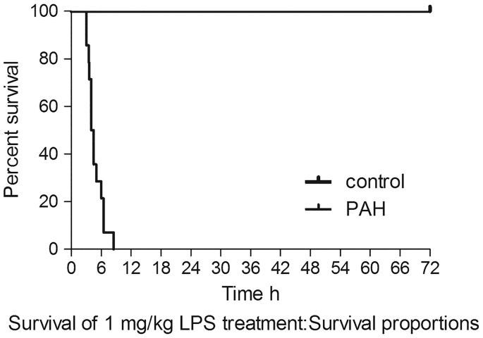
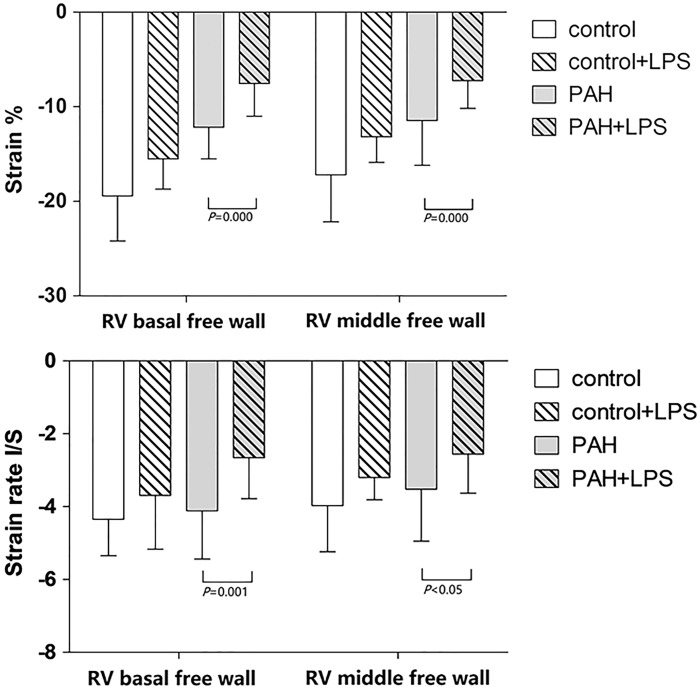
Worsening right ventricular (RV) dysfunction in the presence of pulmonary artery hypertension (PAH) increases morbidity and mortality in this patient population. Transthoracic echocardiography (TTE) is a non-invasive modality to evaluate RV function over time. Using a monocrotaline-induced PAH rat model, we evaluated the effect of acute inflammation on RV function. In this study, both PAH and control rats were injected with Escherichia coli lipopolysaccharide (LPS) to induce an acute inflammatory state. We evaluated survival curves, TTE parameters, and inflammatory markers to better understand the mechanism and impact of acute inflammation on RV function in the presence of PAH. The survival curve of the PAH rats dropped sharply within 9 h after LPS treatment. Several echocardiographic parameters including left ventricular (LV) stroke volume, RV tricuspid annular plane systolic excursion, RV longitudinal peak systolic strain, and strain rate decreased significantly in PAH rats before LPS injection and 2 h after LPS injection. The expression of phospholamban (PLB) and tumor necrosis factor-α (TNF-α) significantly increased and the expression of SERCA2a significantly decreased in PAH rats after LPS administration. LPS suppressed the RV longitudinal peak systolic strain and strain rate and cardiac function deteriorated in PAH rats. These effects may be associated with the signal pathway activity of SERCA2a/PLB.
在肺动脉高压(PAH)患者中,右心室(RV)功能恶化会增加其发病率和死亡率。经胸超声心动图(TTE)是一种用于长期评估RV功能的非侵入性方法。我们使用一种野百合碱诱导的PAH大鼠模型,评估了急性炎症对RV功能的影响。在本研究中,对PAH大鼠和对照大鼠均注射大肠杆菌脂多糖(LPS)以诱导急性炎症状态。我们评估了生存曲线、TTE参数和炎症标志物,以更好地了解急性炎症在PAH存在时对RV功能的机制和影响。PAH大鼠在LPS治疗后9小时内生存曲线急剧下降。包括左心室(LV)每搏输出量、RV三尖瓣环平面收缩期位移、RV纵向峰值收缩应变和应变率在内的几个超声心动图参数在PAH大鼠注射LPS前和注射后2小时显著降低。LPS给药后,PAH大鼠中受磷蛋白(PLB)和肿瘤坏死因子-α(TNF-α)的表达显著增加,而肌浆网钙ATP酶2a(SERCA2a)的表达显著降低。LPS抑制了PAH大鼠的RV纵向峰值收缩应变和应变率,心脏功能恶化。这些影响可能与SERCA2a/PLB的信号通路活性有关。