Larsen Karen B
Department of Pathology, Rigshospitalet, University Hospital of Copenhagen, Copenhagen, Denmark.
Department of Neuropathology and Ocular Pathology, John Radcliffe Hospital, Oxford University Hospital, Oxford, United Kingdom.
Front Neuroanat. 2017 Dec 4;11:112. doi: 10.3389/fnana.2017.00112. eCollection 2017.
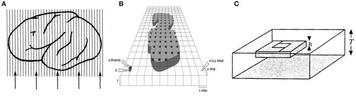
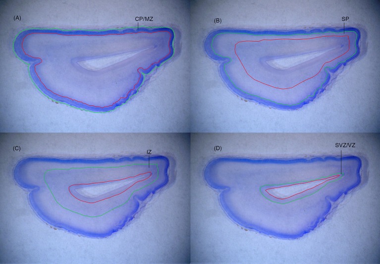
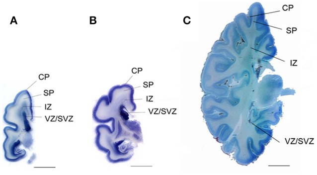
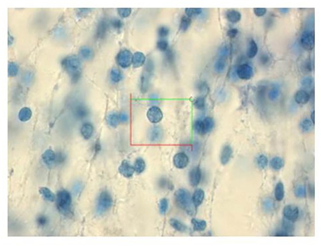
Human fetal brain development is a complex process which is vulnerable to disruption at many stages. Although histogenesis is well-documented, only a few studies have quantified cell numbers across normal human fetal brain growth. Due to the present lack of normative data it is difficult to gauge abnormal development. Furthermore, many studies of brain cell numbers have employed biased counting methods, whereas innovations in stereology during the past 20-30 years enable reliable and efficient estimates of cell numbers. However, estimates of cell volumes and densities in fetal brain samples are unreliable due to unpredictable shrinking artifacts, and the fragility of the fetal brain requires particular care in handling and processing. The optical fractionator design offers a direct and robust estimate of total cell numbers in the fetal brain with a minimum of handling of the tissue. Bearing this in mind, we have used the optical fractionator to quantify the growth of total cell numbers as a function of fetal age. We discovered a two-phased development in total cell numbers in the human fetal forebrain consisting of an initial steep rise in total cell numbers between 13 and 20 weeks of gestation, followed by a slower linear phase extending from mid-gestation to 40 weeks of gestation. Furthermore, we have demonstrated a reduced total cell number in the forebrain in fetuses with Down syndome at midgestation and in intrauterine growth-restricted fetuses during the third trimester.
人类胎儿大脑发育是一个复杂的过程,在许多阶段都容易受到干扰。尽管组织发生已有充分记录,但只有少数研究对正常人类胎儿大脑生长过程中的细胞数量进行了量化。由于目前缺乏标准化数据,很难判断发育是否异常。此外,许多关于脑细胞数量的研究采用了有偏差的计数方法,而在过去20至30年中,体视学的创新使得对细胞数量进行可靠而有效的估计成为可能。然而,由于不可预测的收缩假象,胎儿脑样本中细胞体积和密度的估计并不可靠,而且胎儿脑的脆弱性要求在处理和加工时格外小心。光学分数法设计能够在对组织进行最少处理的情况下,直接且可靠地估计胎儿脑中的总细胞数量。考虑到这一点,我们使用光学分数法来量化总细胞数量随胎龄的增长情况。我们发现人类胎儿前脑的总细胞数量呈两阶段发育,在妊娠13至20周期间总细胞数量最初急剧上升,随后是一个从妊娠中期到40周的较慢线性阶段。此外,我们还证明,患有唐氏综合征的胎儿在妊娠中期以及孕晚期宫内生长受限的胎儿,其前脑的总细胞数量减少。