Neurophysiological Imaging and Modeling Laboratory, University of Idaho, 875 Perimeter Dr. MC1122, Moscow, ID, 83844-1122, USA.
Seattle Science Foundation, 200 2nd Ave N, Seattle, WA, 98109, USA.
Fluids Barriers CNS. 2017 Dec 19;14(1):36. doi: 10.1186/s12987-017-0085-y.
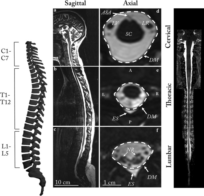
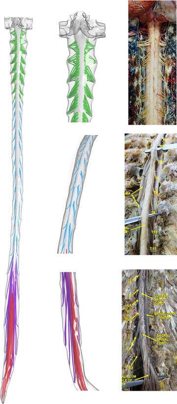
The spinal subarachnoid space (SSS) has a complex 3D fluid-filled geometry with multiple levels of anatomic complexity, the most salient features being the spinal cord and dorsal and ventral nerve rootlets. An accurate anthropomorphic representation of these features is needed for development of in vitro and numerical models of cerebrospinal fluid (CSF) dynamics that can be used to inform and optimize CSF-based therapeutics.
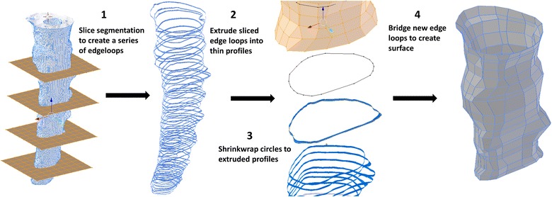
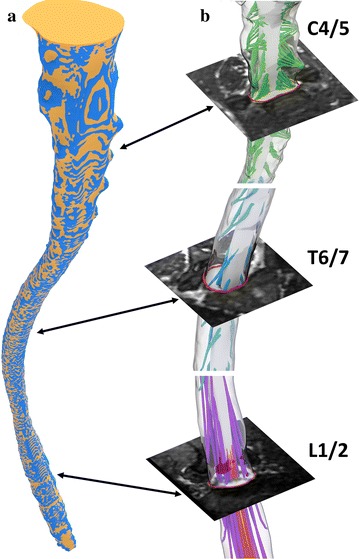
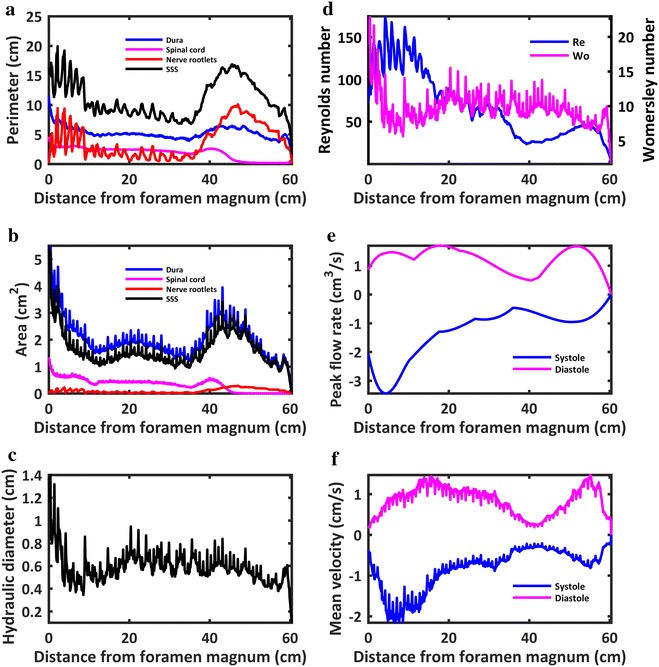
A subject-specific 3D model of the SSS was constructed based on high-resolution anatomic MRI. An expert operator completed manual segmentation of the CSF space with detailed consideration of the anatomy. 31 pairs of semi-idealized dorsal and ventral nerve rootlets (NR) were added to the model based on anatomic reference to the magnetic resonance (MR) imaging and cadaveric measurements in the literature. Key design criteria for each NR pair included the radicular line, descending angle, number of NR, attachment location along the spinal cord and exit through the dura mater. Model simplification and smoothing was performed to produce a final model with minimum vertices while maintaining minimum error between the original segmentation and final design. Final model geometry and hydrodynamics were characterized in terms of axial distribution of Reynolds number, Womersley number, hydraulic diameter, cross-sectional area and perimeter.
The final model had a total of 139,901 vertices with a total CSF volume within the SSS of 97.3 cm. Volume of the dura mater, spinal cord and NR was 123.1, 19.9 and 5.8 cm. Surface area of these features was 318.52, 112.2 and 232.1 cm respectively. Maximum Reynolds number was 174.9 and average Womersley number was 9.6, likely indicating presence of a laminar inertia-dominated oscillatory CSF flow field.
This study details an anatomically realistic anthropomorphic 3D model of the SSS based on high-resolution MR imaging of a healthy human adult female. The model is provided for re-use under the Creative Commons Attribution-ShareAlike 4.0 International license (CC BY-SA 4.0) and can be used as a tool for development of in vitro and numerical models of CSF dynamics for design and optimization of intrathecal therapeutics.
脊髓蛛网膜下腔(SSS)具有复杂的三维充满液体的几何形状,具有多个解剖学复杂层次,最显著的特征是脊髓和背根与腹根。为了开发可以用于告知和优化基于脑脊液的治疗方法的体外和数值脑脊液动力学模型,需要对这些特征进行准确的拟人化表示。
基于高分辨率解剖磁共振成像构建了 SSS 的特定于个体的 3D 模型。一位专家操作员使用详细的解剖考虑完成了 CSF 空间的手动分割。根据磁共振成像(MR)成像和文献中的尸体测量结果,在模型中添加了 31 对半理想的背根和腹根(NR)。每对 NR 的关键设计标准包括神经根线、下降角度、NR 的数量、沿脊髓的附着位置以及通过硬脑膜的出口。对模型进行简化和平滑处理,生成具有最小顶点的最终模型,同时保持原始分割和最终设计之间的最小误差。最终模型的几何形状和流体动力学特性采用轴向雷诺数、沃默斯利数、水力直径、横截面积和周长的分布来描述。
最终模型共有 139901 个顶点,SSS 内的脑脊液总量为 97.3cm。硬脑膜、脊髓和 NR 的体积分别为 123.1cm、19.9cm 和 5.8cm。这些特征的表面积分别为 318.52cm、112.2cm 和 232.1cm。最大雷诺数为 174.9,平均沃默斯利数为 9.6,这可能表明存在层流惯性主导的振荡脑脊液流场。
本研究详细介绍了一种基于健康成年女性高分辨率磁共振成像的 SSS 的解剖逼真的拟人化 3D 模型。该模型根据知识共享署名-相同方式共享 4.0 国际许可证(CC BY-SA 4.0)提供再使用,并可作为体外和数值脑脊液动力学模型的开发工具,用于设计和优化鞘内治疗。