Rocha Sara, Freitas Ana, Guimaraes Sofia C, Vitorino Rui, Aroso Miguel, Gomez-Lazaro Maria
i3S-Instituto de Investigação e Inovação em Saúde, Universidade do Porto, 4200-135 Porto, Portugal.
IBMC-Instituto de Biologia Molecular e Celular, Universidade do Porto, 4200-135 Porto, Portugal.
Antioxidants (Basel). 2017 Dec 21;7(1):1. doi: 10.3390/antiox7010001.
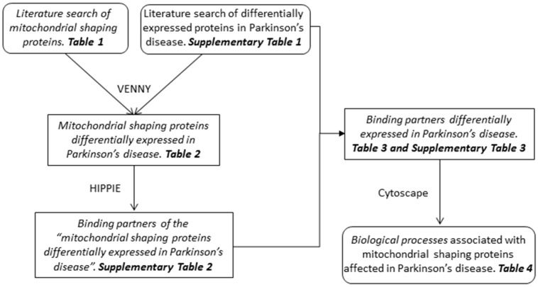
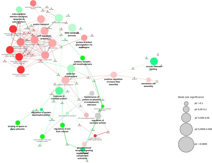
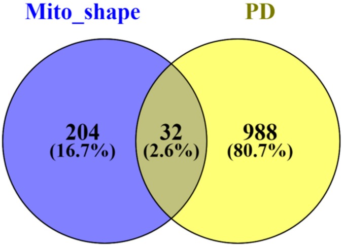
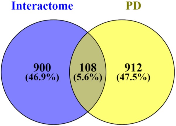
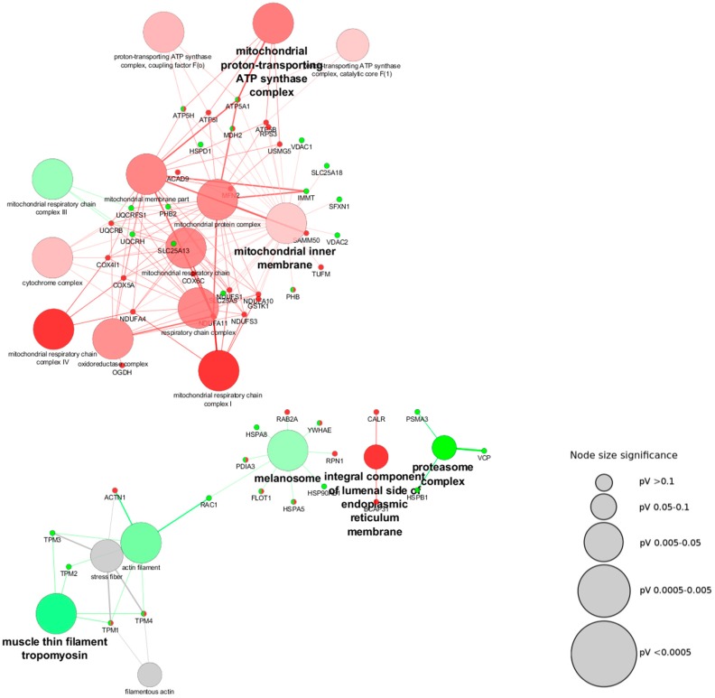
It has long been accepted that mitochondrial function and morphology is affected in Parkinson's disease, and that mitochondrial function can be directly related to its morphology. So far, mitochondrial morphological alterations studies, in the context of this neurodegenerative disease, have been performed through microscopic methodologies. The goal of the present work is to address if the modifications in the mitochondrial-shaping proteins occurring in this disorder have implications in other cellular pathways, which might constitute important pathways for the disease progression. To do so, we conducted a novel approach through a thorough exploration of the available proteomics-based studies in the context of Parkinson's disease. The analysis provided insight into the altered biological pathways affected by changes in the expression of mitochondrial-shaping proteins via different bioinformatic tools. Unexpectedly, we observed that the mitochondrial-shaping proteins altered in the context of Parkinson's disease are, in the vast majority, related to the organization of the mitochondrial cristae. Conversely, in the studies that have resorted to microscopy-based techniques, the most widely reported alteration in the context of this disorder is mitochondria fragmentation. Cristae membrane organization is pivotal for mitochondrial ATP production, and changes in their morphology have a direct impact on the organization and function of the oxidative phosphorylation (OXPHOS) complexes. To understand which biological processes are affected by the alteration of these proteins we analyzed the binding partners of the mitochondrial-shaping proteins that were found altered in Parkinson's disease. We showed that the binding partners fall into seven different cellular components, which include mitochondria, proteasome, and endoplasmic reticulum (ER), amongst others. It is noteworthy that, by evaluating the biological process in which these modified proteins are involved, we showed that they are related to the production and metabolism of ATP, immune response, cytoskeleton alteration, and oxidative stress, amongst others. In summary, with our bioinformatics approach using the data on the modified proteins in Parkinson's disease patients, we were able to relate the alteration of mitochondrial-shaping proteins to modifications of crucial cellular pathways affected in this disease.
长期以来,人们一直认为帕金森病中线粒体功能和形态会受到影响,且线粒体功能可能与其形态直接相关。到目前为止,在这种神经退行性疾病背景下进行的线粒体形态改变研究,都是通过显微镜方法进行的。本研究的目的是探讨这种疾病中发生的线粒体塑形蛋白修饰是否会对其他细胞途径产生影响,而这些途径可能构成疾病进展的重要途径。为此,我们通过全面探索帕金森病背景下基于蛋白质组学的现有研究,采用了一种新方法。通过不同的生物信息学工具,该分析深入了解了受线粒体塑形蛋白表达变化影响的改变的生物途径。出乎意料的是,我们观察到在帕金森病背景下发生改变的线粒体塑形蛋白,绝大多数与线粒体嵴组织有关。相反,在采用基于显微镜技术的研究中,该疾病背景下报道最多的改变是线粒体碎片化。嵴膜组织对于线粒体ATP生成至关重要,其形态变化对氧化磷酸化(OXPHOS)复合物的组织和功能有直接影响。为了了解哪些生物过程受这些蛋白质改变的影响,我们分析了在帕金森病中发现发生改变的线粒体塑形蛋白的结合伙伴。我们发现这些结合伙伴分为七个不同的细胞成分,包括线粒体、蛋白酶体和内质网等。值得注意的是,通过评估这些修饰蛋白所涉及的生物过程,我们发现它们与ATP的产生和代谢、免疫反应、细胞骨架改变以及氧化应激等有关。总之,通过我们利用帕金森病患者修饰蛋白数据的生物信息学方法,我们能够将线粒体塑形蛋白的改变与该疾病中受影响的关键细胞途径的修饰联系起来。