Noble Jonathan J, Chruscikowski Emily, Fry Nicola R D, Lewis Andrew P, Gough Martin, Shortland Adam P
One Small Step Gait Laboratory, Evelina London Children's Hospital, Guy's and St Thomas' NHS Foundation Trust, Guy's Hospital, London, SE1 9RT, UK.
Division of Imaging Sciences and Biomedical Engineering, King's College London, The Rayne Institute, 4th Floor, Lambeth Wing, St Thomas' Hospital, London, SE1 7EH, UK.
BMC Neurol. 2017 Dec 29;17(1):223. doi: 10.1186/s12883-017-1005-0.
Individuals with cerebral palsy have smaller muscle volumes normalised to body mass than their typically developing peers. The aim of this study is to investigate the relationship between lower limb muscle volume and body mass in young people with bilateral cerebral palsy and their typically developing peers.
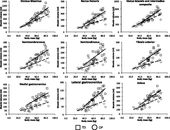
Twenty-five participants with bilateral cerebral palsy (aged 14.7±3.0 years, GMFCS level I-III) and 25 of their typically developing peers (aged 16.8±3.3 years) took part in this study. None of the participants had undergone orthopaedic surgery, botulinum toxin injections, or serial casting in the previous year. All participants underwent magnetic resonance imaging of both lower limbs. Nine major muscles of each lower limb were individually manually segmented and the muscle volumes calculated.
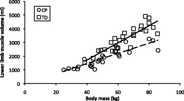
Body mass and total lower limb muscle volume were significantly linearly related in both the cerebral palsy (R = 0.75, p<0.001) and typically developing (R = 0.77, p<0.001) groups. The slope of the relationship between muscle volume and body mass was significantly shallower in the cerebral palsy group compared to the typically developing group (p=0.007).
This cross-sectional study suggests that the increase in size of lower limb muscles relative to body mass is reduced in adolescents and young adults with cerebral palsy. Longitudinal studies are required to further investigate altered muscle growth trajectories in this group and their impact on long-term mobility.
与发育正常的同龄人相比,脑性瘫痪患者经体重标准化后的肌肉体积更小。本研究旨在调查双侧脑性瘫痪青少年及其发育正常的同龄人下肢肌肉体积与体重之间的关系。
25名双侧脑性瘫痪患者(年龄14.7±3.0岁,GMFCS分级I - III级)及其25名发育正常的同龄人(年龄16.8±3.3岁)参与了本研究。所有参与者在前一年均未接受过骨科手术、肉毒杆菌毒素注射或连续石膏固定治疗。所有参与者均接受了双下肢的磁共振成像检查。对每个下肢的九块主要肌肉进行单独手动分割并计算肌肉体积。
脑性瘫痪组(R = 0.75,p<0.001)和发育正常组(R = 0.77,p<0.001)的体重与下肢总肌肉体积均呈显著线性相关。与发育正常组相比,脑性瘫痪组肌肉体积与体重之间关系的斜率明显更平缓(p = 0.007)。
这项横断面研究表明,患有脑性瘫痪的青少年和年轻人下肢肌肉相对于体重的增长有所减少。需要进行纵向研究以进一步调查该组肌肉生长轨迹的改变及其对长期活动能力的影响。