Daviet-Noual Victor, Ejeil Anne-Laure, Gossiome Charles, Moreau Nathan, Salmon Benjamin
Dental Medicine Department, Bretonneau Hospital, HUPNVS, AP-HP, Paris, France.
Laboratory of Orofacial Neurobiology - Paris Diderot University, Paris, France.
BMC Oral Health. 2017 Dec 28;17(1):161. doi: 10.1186/s12903-017-0455-5.
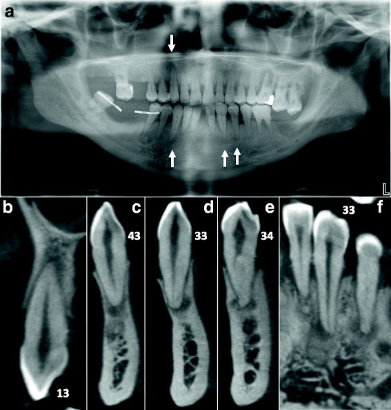
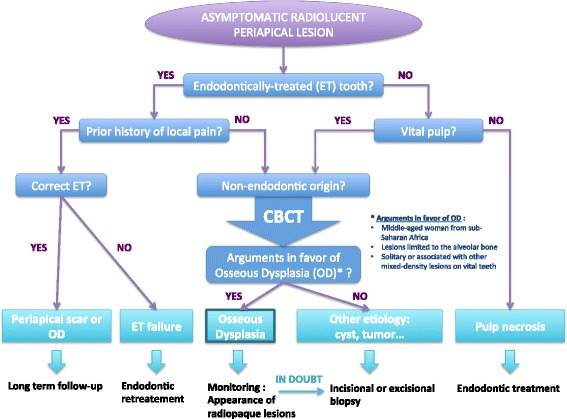
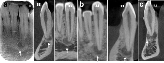
Osseous dysplasia (OD) is the most common fibro-osseous lesion of the jaw affecting the periapical region. Early stages of OD can resemble periapical radiolucencies, thus mimicking the radiological aspects of an endodontic pathology. Such radiolucent lesions affecting previously decayed or treated teeth are even more complex to interpret.
The aim of this paper is to report a case-series of representative clinical situations describing the radiological features and illustrating the diagnostic workup of patients with florid osseous dysplasia (FOD). Emphasis is given to the endodontic implications of such periapical bone disease and the complexity of accurate diagnosis in the context of endodontic retreatment. We then propose a practical radiological-based diagnostic algorithm to assist the clinician in the diagnostic of OD periapical lesions.
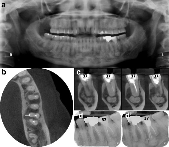
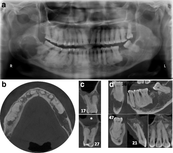
Periapical lesions may be confused with bone diseases such as osseous dysplasia, especially in the radiolucent initial stage. Knowledge of clinical features associated with a careful reading of cone beam CT images, such as fine opacities within the hypodense periapical lesion, may help determine the right diagnostic.
骨发育异常(OD)是颌骨最常见的纤维-骨病变,累及根尖区。OD的早期阶段可类似于根尖周透射影,从而模仿牙髓病的影像学表现。这种影响先前已龋坏或已治疗牙齿的透射性病变更难以解释。
本文旨在报告一系列具有代表性的临床情况,描述其影像学特征,并说明 florid 骨发育异常(FOD)患者的诊断检查。重点关注此类根尖周骨病的牙髓病学意义以及在牙髓再治疗背景下准确诊断的复杂性。然后,我们提出一种基于影像学的实用诊断算法,以协助临床医生诊断OD根尖周病变。
根尖周病变可能与骨发育异常等骨病相混淆,尤其是在透射性初始阶段。了解与仔细阅读锥形束CT图像相关的临床特征,如低密度根尖周病变内的细微不透光区,可能有助于做出正确诊断。