Boitor Radu, Kong Kenny, Shipp Dustin, Varma Sandeep, Koloydenko Alexey, Kulkarni Kusum, Elsheikh Somaia, Schut Tom Bakker, Caspers Peter, Puppels Gerwin, van der Wolf Martin, Sokolova Elena, Nijsten T E C, Salence Brogan, Williams Hywel, Notingher Ioan
School of Physics and Astronomy, University Park, University of Nottingham, Nottingham, NG7 2RD, UK.
Circle Nottingham Ltd NHS Treatment Centre, Lister Road, Nottingham NG7 2FT, UK.
Biomed Opt Express. 2017 Nov 22;8(12):5749-5766. doi: 10.1364/BOE.8.005749. eCollection 2017 Dec 1.
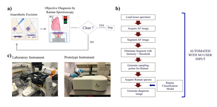
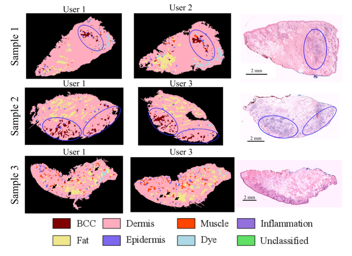
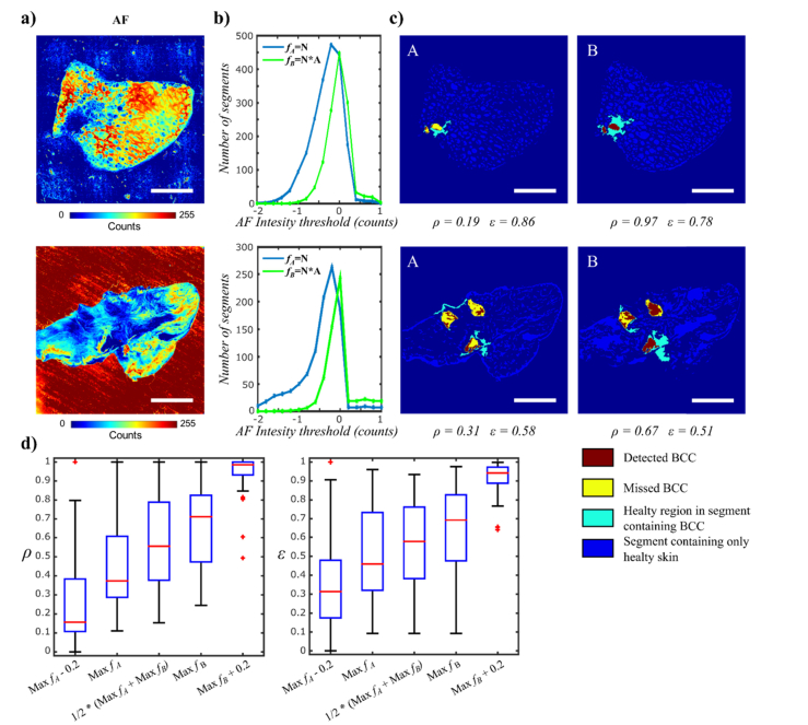
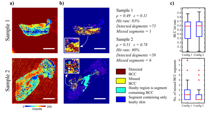
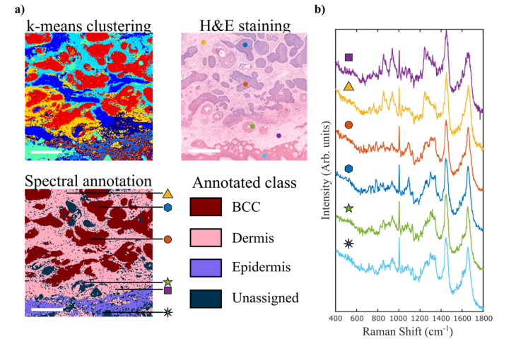
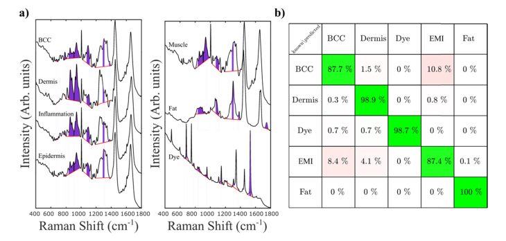
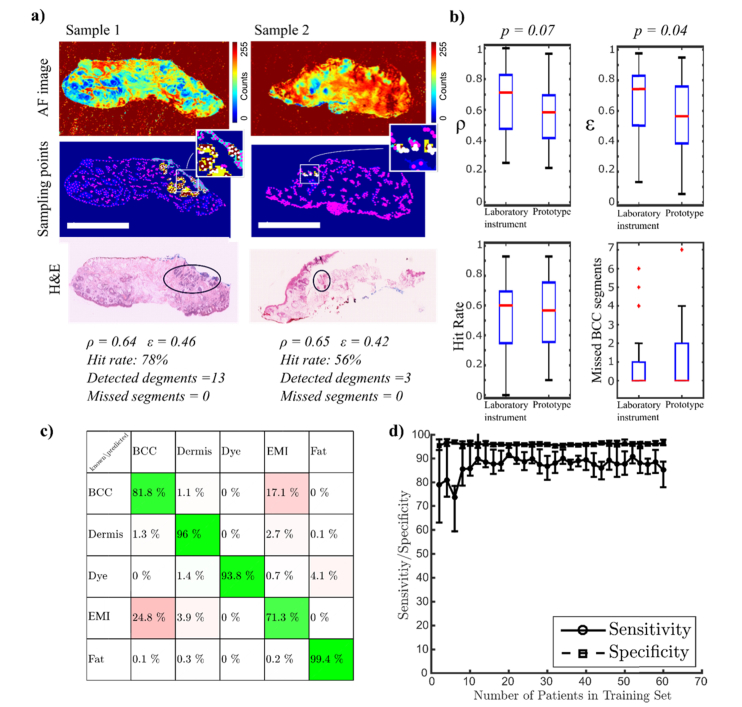
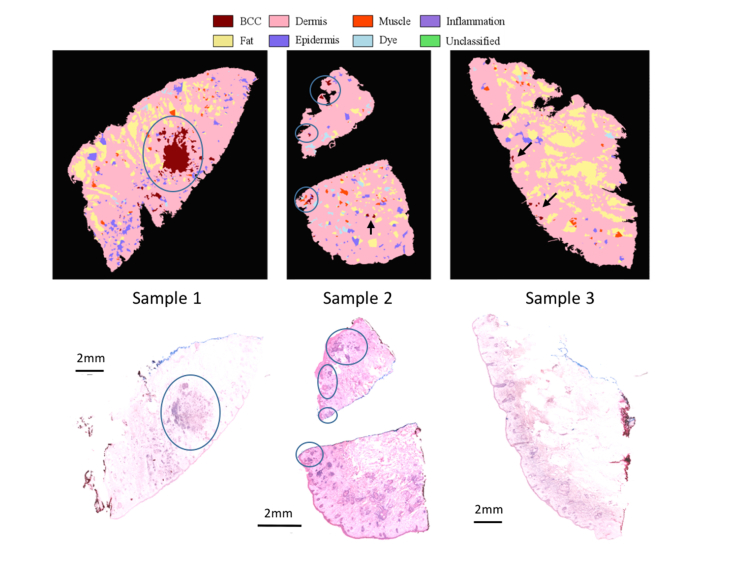
Multimodal spectral histopathology (MSH), an optical technique combining tissue auto-fluorescence (AF) imaging and Raman micro-spectroscopy (RMS), was previously proposed for detection of residual basal cell carcinoma (BCC) at the surface of surgically-resected skin tissue. Here we report the development of a fully-automated prototype instrument based on MSH designed to be used in the clinic and operated by a non-specialist spectroscopy user. The algorithms for the AF image processing and Raman spectroscopy classification had been first optimised on a manually-operated laboratory instrument and then validated on the automated prototype using skin samples from independent patients. We present results on a range of skin samples excised during Mohs micrographic surgery, and demonstrate consistent diagnosis obtained in repeat test measurement, in agreement with the reference histopathology diagnosis. We also show that the prototype instrument can be operated by clinical users (a skin surgeon and a core medical trainee, after only 1-8 hours of training) to obtain consistent results in agreement with histopathology. The development of the new automated prototype and demonstration of inter-instrument transferability of the diagnosis models are important steps on the clinical translation path: it allows the testing of the MSH technology in a relevant clinical environment in order to evaluate its performance on a sufficiently large number of patients.
多模态光谱组织病理学(MSH)是一种将组织自体荧光(AF)成像与拉曼显微光谱(RMS)相结合的光学技术,此前已被提出用于检测手术切除的皮肤组织表面残留的基底细胞癌(BCC)。在此,我们报告了一种基于MSH的全自动原型仪器的开发情况,该仪器设计用于临床,由非专业光谱学用户操作。AF图像处理和拉曼光谱分类算法首先在手动操作的实验室仪器上进行了优化,然后使用独立患者的皮肤样本在自动原型上进行了验证。我们展示了在莫氏显微外科手术期间切除的一系列皮肤样本的结果,并证明了在重复测试测量中获得的一致诊断结果,与参考组织病理学诊断结果相符。我们还表明,临床用户(一名皮肤外科医生和一名核心医学实习生,仅经过1 - 8小时的培训)可以操作该原型仪器,以获得与组织病理学相符的一致结果。新型自动原型的开发以及诊断模型的仪器间可转移性的证明是临床转化道路上的重要步骤:它允许在相关临床环境中测试MSH技术,以便在足够多的患者身上评估其性能。