Division of Cardiothoracic Surgery, Cardiovascular Research Center, Rhode Island Hospital, Alpert Medical School of Brown University, Providence, RI, USA.
Cardiothoracic Surgery Research Laboratory, Rhode Island Hospital, 1 Hoppin Street, Coro West Room 5.229, Providence, RI, 02903, USA.
Mol Cell Biochem. 2018 Aug;445(1-2):187-194. doi: 10.1007/s11010-017-3264-x. Epub 2018 Jan 5.
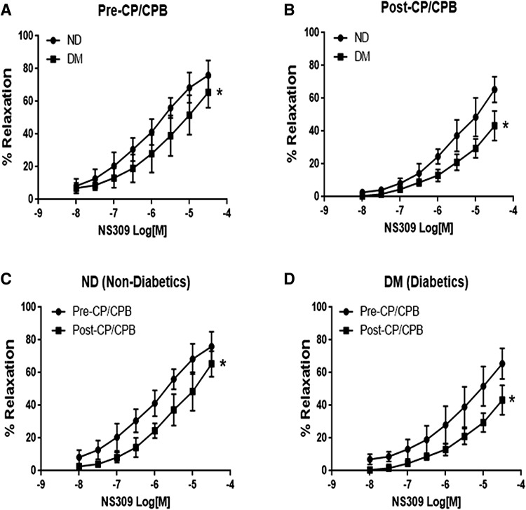
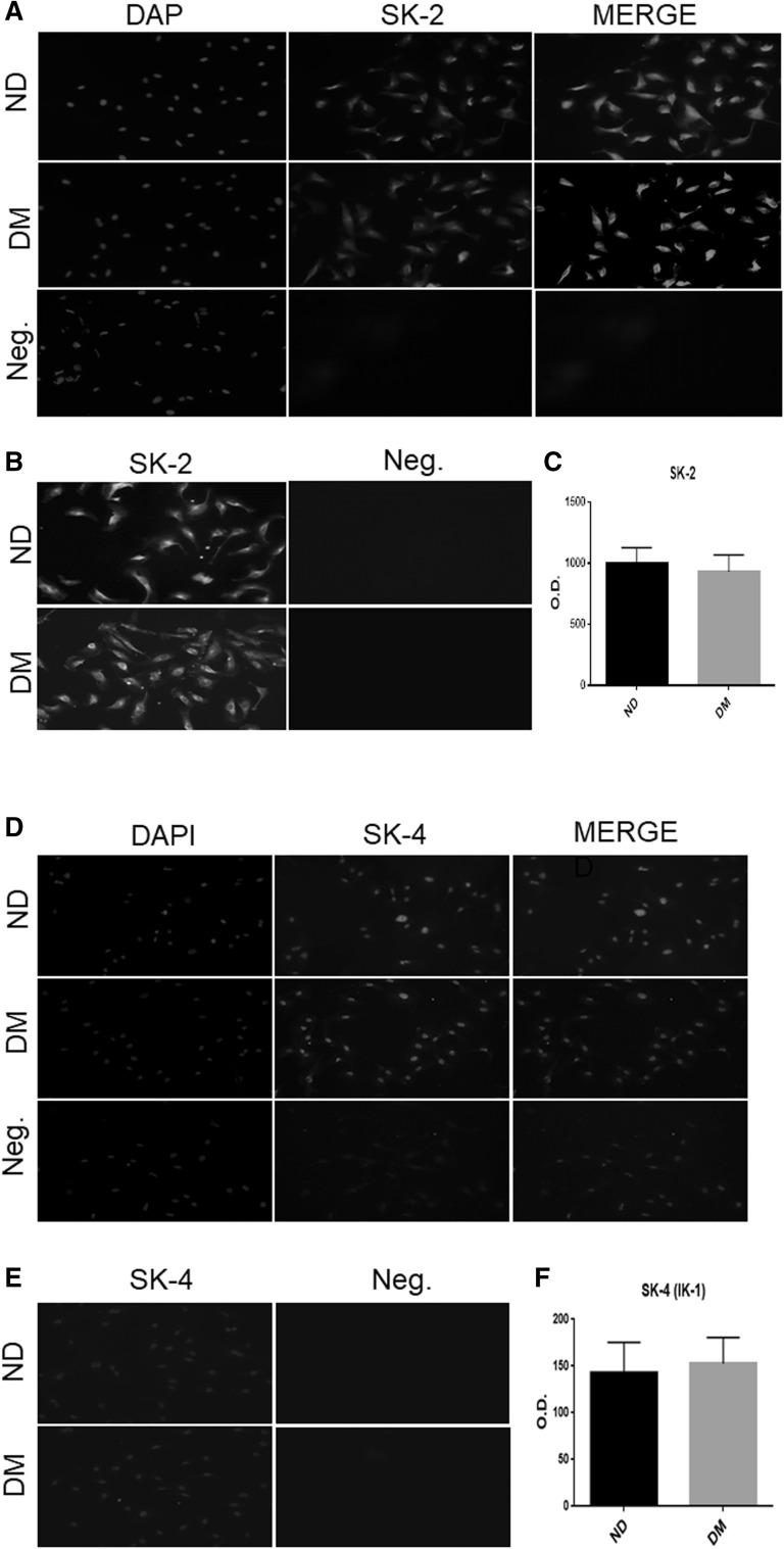
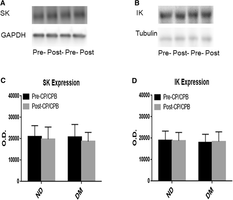
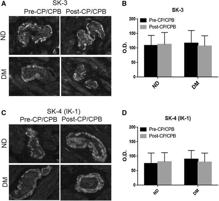
We have recently found that diabetes is associated with the inactivation of the calcium-activated potassium channels (K) in endothelial cells, which may contribute to endothelial dysfunction in diabetic patients at baseline. In the current study, we further investigated the effects of diabetes on coronary arteriolar responses to the small (SK) and intermediate (IK) K opener NS309 in diabetic and non-diabetic patients and correlated that data with the changes in the SK/IK protein expression/distribution in the setting of cardioplegic ischemia and reperfusion (CP) and cardiopulmonary bypass (CPB). Coronary arterioles from the harvested right atrial tissue samples from diabetic and non-diabetic patients (n = 8/group) undergoing cardiac surgery were dissected pre- and post-CP/CPB. The in vitro relaxation response of pre-contracted arterioles was examined in the presence of the selective SK/IK opener NS309 (10-10 M). The protein expression/localization of K channels in the harvested atrial tissue samples, coronary microvessels, and primary cultured human coronary endothelial cells were assayed by Western blotting and immunohistochemistry. The relaxation response to NS309 post-CP/CPB was significantly decreased in diabetic and non-diabetic groups compared to their pre-CP/CPB responses, respectively (P < 0.05). Furthermore, this decrease was greater in the diabetic group than that of the non-diabetic group (P < 0.05). There were no significant differences in the total protein expression/distribution of SK/IK in the human myocardium, coronary microvessels or coronary endothelial cells between diabetic and non-diabetic groups or between pre- and post-CP/CPB (P > 0.05). Our results suggest that diabetes further inactivates SK/IK channels of coronary microvasculature early after CP/CPB and cardiac surgery. The lack of diabetic changes in SK/IK protein abundances in the setting of CP/CPB suggests that the effect is post-translational. This alteration may contribute to post-operative endothelial dysfunction in the diabetic patients early after CP/CPB and cardiac surgery.
我们最近发现,糖尿病会导致内皮细胞中钙激活钾通道(K 通道)失活,这可能导致糖尿病患者在基线时内皮功能障碍。在目前的研究中,我们进一步研究了糖尿病对糖尿病和非糖尿病患者冠状小动脉(SK)和中间(IK)K 开放剂 NS309 反应的影响,并将数据与心脏停搏(CP)和体外循环(CPB)期间 SK/IK 蛋白表达/分布的变化相关联。来自接受心脏手术的糖尿病和非糖尿病患者(每组 n=8)的右心房组织样本中分离出冠状小动脉,并在 CP/CPB 前后进行解剖。在存在选择性 SK/IK 开放剂 NS309(10-10 M)的情况下,检查预先收缩的小动脉的体外松弛反应。通过 Western blot 和免疫组织化学检测收获的心房组织样本、冠状微血管和原代培养的人冠状动脉内皮细胞中的 K 通道蛋白表达/定位。与 CP/CPB 前反应相比,CP/CPB 后 NS309 的松弛反应在糖尿病和非糖尿病组中均显著降低(P < 0.05)。此外,糖尿病组的这种下降大于非糖尿病组(P < 0.05)。在糖尿病和非糖尿病组之间或 CP/CPB 前后,人心肌、冠状微血管或冠状内皮细胞中 SK/IK 的总蛋白表达/分布均无显著差异(P > 0.05)。我们的结果表明,糖尿病在 CP/CPB 和心脏手术后早期进一步使冠状微血管的 SK/IK 通道失活。CP/CPB 期间 SK/IK 蛋白丰度无糖尿病变化表明该效应是翻译后发生的。这种改变可能导致 CP/CPB 和心脏手术后早期糖尿病患者的术后内皮功能障碍。