• 文献检索
  • 文档翻译
  • 深度研究
  • 学术资讯
  • Suppr Zotero 插件Zotero 插件
  • 邀请有礼
  • 套餐&价格
  • 历史记录
应用&插件
Suppr Zotero 插件Zotero 插件浏览器插件Mac 客户端Windows 客户端微信小程序
定价
高级版会员购买积分包购买API积分包
服务
文献检索文档翻译深度研究API 文档MCP 服务
关于我们
关于 Suppr公司介绍联系我们用户协议隐私条款
关注我们

Suppr 超能文献

核心技术专利:CN118964589B侵权必究
粤ICP备2023148730 号-1Suppr @ 2026

文献检索

告别复杂PubMed语法,用中文像聊天一样搜索,搜遍4000万医学文献。AI智能推荐,让科研检索更轻松。

立即免费搜索

文件翻译

保留排版,准确专业,支持PDF/Word/PPT等文件格式,支持 12+语言互译。

免费翻译文档

深度研究

AI帮你快速写综述,25分钟生成高质量综述,智能提取关键信息,辅助科研写作。

立即免费体验

钆特酸葡胺会根据 b 值组合降低乳腺病变的 ADC 值。

Gadoterate meglumine decreases ADC values of breast lesions depending on the b value combination.

机构信息

Kuopio University Hospital, Diagnostic Imaging Centre, Department of Clinical Radiology, Kuopio University Hospital, PO Box 100, Puijonlaaksontie 2, 70029, Kuopio, Finland.

University of Eastern Finland, Institute of Clinical Medicine, School of Medicine, Department of Clinical Radiology, Kuopio University Hospital, PO Box 1777, Puijonlaaksontie 2, 70210, Kuopio, Finland.

出版信息

Sci Rep. 2018 Jan 8;8(1):87. doi: 10.1038/s41598-017-18035-0.

DOI:10.1038/s41598-017-18035-0
PMID:29311709
原文链接:https://pmc.ncbi.nlm.nih.gov/articles/PMC5758819/
Abstract

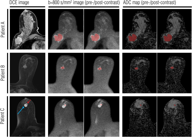
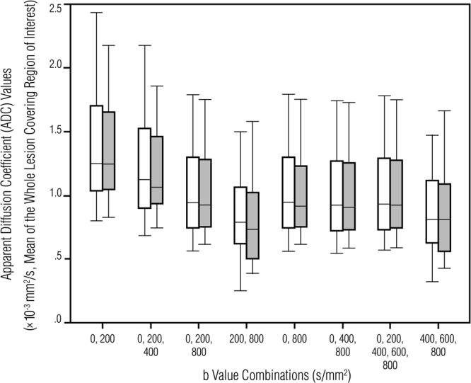
To retrospectively evaluated the influence of administration of the gadolinium based intravenous contrast agent (G-CA) on apparent diffusion coefficient (ADC) values in ADC maps generated using multiple b value combinations. A total of 106 women underwent bilateral 3.0 T breast MRI. As an internal validation, diffusion-weighted imaging (b values of 0, 200, 400, 600, 800 s/mm) was performed before and after the G-CA (gadoterate meglumine (0.2 ml/kg, 3 ml/s)). Whole lesion and fibroglandular tissue (FGT) covering region-of-interests (ROIs) were drawn on the b = 800 s/mm images; ROIs were then propagated to multiple retrospectively generated ADC maps. Twenty-seven patients (mean age 55.8 ± 10.8 years) with 32 mass-like enhancing breast lesions including 25 (78.1 %) histopathologically malignant lesions were enrolled. Lesion ADC values were statistically significantly higher in pre-G-CA than post-G-CA ADC maps (ADC: 1.05 ± 0.35 × 10 mm/s vs. 1.02 ± 0.36 × 10 mm/s (P < 0.05); ADC: 1.25 ± 0.42 × 10 mm/s vs. 1.20 ± 0.35 × 10 mm/s (P < 0.05)). ADC values between pre- and post-contrast maps were not statistically different when the maps were generated using other b value combinations. Contrast agent administration did not affect the FGT ADC values. G-CA statistically significantly reduced the ADC values of breast lesions on ADC maps generated using the clinically widely utilized b values.

摘要

目的

回顾性评估在使用不同 b 值组合生成 ADC 图时,静脉注射钆基对比剂(G-CA)给药对表观扩散系数(ADC)值的影响。

方法

共 106 名女性接受双侧 3.0T 乳腺 MRI 检查。作为内部验证,在 G-CA(钆特酸葡胺(0.2ml/kg,3ml/s))给药前后进行扩散加权成像(b 值为 0、200、400、600、800s/mm)。在 b=800s/mm 图像上对整个病变和纤维腺体组织(FGT)覆盖 ROI 进行勾画;然后将 ROI 传播到多个回顾性生成的 ADC 图中。

结果

共纳入 27 例(平均年龄 55.8±10.8 岁)、32 个肿块样强化乳腺病变患者,其中 25 例(78.1%)经病理证实为恶性病变。与 G-CA 后 ADC 图相比,G-CA 前 ADC 图的病变 ADC 值明显升高(ADC:1.05±0.35×10mm/s 比 1.02±0.36×10mm/s(P<0.05);ADC:1.25±0.42×10mm/s 比 1.20±0.35×10mm/s(P<0.05))。当使用其他 b 值组合生成 ADC 图时,前后对比图的 ADC 值无统计学差异。对比剂给药未影响 FGT 的 ADC 值。G-CA 给药后,使用临床广泛应用的 b 值生成的 ADC 图中,乳腺病变的 ADC 值明显降低。

结论

G-CA 给药后,使用临床广泛应用的 b 值生成的 ADC 图中,乳腺病变的 ADC 值明显降低。

https://cdn.ncbi.nlm.nih.gov/pmc/blobs/d3ea/5758819/cc9e9c9b2aa0/41598_2017_18035_Fig2_HTML.jpg
https://cdn.ncbi.nlm.nih.gov/pmc/blobs/d3ea/5758819/3cad477f7004/41598_2017_18035_Fig1_HTML.jpg
https://cdn.ncbi.nlm.nih.gov/pmc/blobs/d3ea/5758819/cc9e9c9b2aa0/41598_2017_18035_Fig2_HTML.jpg
https://cdn.ncbi.nlm.nih.gov/pmc/blobs/d3ea/5758819/3cad477f7004/41598_2017_18035_Fig1_HTML.jpg
https://cdn.ncbi.nlm.nih.gov/pmc/blobs/d3ea/5758819/cc9e9c9b2aa0/41598_2017_18035_Fig2_HTML.jpg

相似文献

1
Gadoterate meglumine decreases ADC values of breast lesions depending on the b value combination.钆特酸葡胺会根据 b 值组合降低乳腺病变的 ADC 值。
Sci Rep. 2018 Jan 8;8(1):87. doi: 10.1038/s41598-017-18035-0.
2
Diffusion-weighted imaging: Effects of intravascular contrast agents on apparent diffusion coefficient measures of breast malignancies at 3 Tesla.扩散加权成像:血管内造影剂对3特斯拉下乳腺恶性肿瘤表观扩散系数测量值的影响。
J Magn Reson Imaging. 2015 Sep;42(3):788-800. doi: 10.1002/jmri.24844. Epub 2015 Jan 22.
3
Improving malignancy prediction in breast lesions with the combination of apparent diffusion coefficient and dynamic contrast-enhanced kinetic descriptors.结合表观扩散系数和动态对比增强动力学描述符提高乳腺病变的恶性预测能力
Clin Radiol. 2015 Sep;70(9):1016-25. doi: 10.1016/j.crad.2015.05.009. Epub 2015 Jun 28.
4
Diffusion-weighted MR imaging of benign and malignant breast lesions before and after contrast enhancement.对比增强前后乳腺良恶性病变的扩散加权磁共振成像
Rofo. 2014 Feb;186(2):130-5. doi: 10.1055/s-0033-1350298. Epub 2013 Aug 8.
5
Effect of gadolinium chelate contrast agents on diffusion weighted MR imaging of the liver, spleen, pancreas and kidney at 3 T.3T 磁共振肝脏、脾脏、胰腺和肾脏弥散加权成像中钆螯合物对比剂的作用。
Eur J Radiol. 2011 Nov;80(2):e1-7. doi: 10.1016/j.ejrad.2010.05.019. Epub 2010 Jun 20.
6
Evaluation of 3.0-T MRI Brain Signal after Exposure to Gadoterate Meglumine in Women with High Breast Cancer Risk and Screening Breast MRI.高乳腺癌风险女性行乳腺 MRI 筛查中钆喷酸葡胺暴露后脑磁共振信号 3.0-T 成像评估
Radiology. 2019 Dec;293(3):523-530. doi: 10.1148/radiol.2019190847. Epub 2019 Oct 22.
7
The diagnostic value of diffusion-weighted imaging and the apparent diffusion coefficient values in the differentiation of benign and malignant breast lesions.扩散加权成像及表观扩散系数值在乳腺良恶性病变鉴别诊断中的价值
J Med Imaging Radiat Oncol. 2015 Apr;59(2):141-8. doi: 10.1111/1754-9485.12273. Epub 2015 Jan 7.
8
Pharmacokinetic approach for dynamic breast MRI to indicate signal intensity time curves of benign and malignant lesions by using the tumor flow residence time.利用肿瘤血流滞留时间对动态乳腺 MRI 的药代动力学方法,对良、恶性病变的信号强度时间曲线进行指示。
Invest Radiol. 2013 Feb;48(2):69-78. doi: 10.1097/RLI.0b013e31827d29cf.
9
Apparent diffusion coefficient mapping using diffusion-weighted MRI: impact of background parenchymal enhancement, amount of fibroglandular tissue and menopausal status on breast cancer diagnosis.使用磁共振扩散加权成像进行表观扩散系数图:背景实质强化、纤维腺体组织含量和绝经状态对乳腺癌诊断的影响。
Eur Radiol. 2018 Jun;28(6):2516-2524. doi: 10.1007/s00330-017-5202-4. Epub 2018 Jan 12.
10
The role of apparent diffusion coefficient values in the differential diagnosis of breast lesions in diffusion-weighted MRI.表观扩散系数值在弥散加权 MRI 鉴别诊断乳腺病变中的作用。
Diagn Interv Radiol. 2013 Nov-Dec;19(6):457-62. doi: 10.5152/dir.2013.12132.

引用本文的文献

1
The relevance of T2 relaxation time in interpreting MRI apparent diffusion coefficient (ADC) map for musculoskeletal structures.T2弛豫时间在解读肌肉骨骼结构的MRI表观扩散系数(ADC)图中的相关性。
Quant Imaging Med Surg. 2023 Dec 1;13(12):7657-7666. doi: 10.21037/qims-23-1392. Epub 2023 Oct 20.
2
Peritumor to tumor apparent diffusion coefficient ratio is associated with biologically more aggressive breast cancer features and correlates with the prognostication tools.肿瘤周围至肿瘤表观扩散系数比值与生物学侵袭性更强的乳腺癌特征相关,并与预后评估工具相关。
PLoS One. 2020 Jun 25;15(6):e0235278. doi: 10.1371/journal.pone.0235278. eCollection 2020.
3

本文引用的文献

1
MR Imaging for Diagnosis of Malignancy in Mammographic Microcalcifications: A Systematic Review and Meta-Analysis.磁共振成像在乳腺微钙化良恶性诊断中的应用:系统评价和荟萃分析。
Radiology. 2017 Jun;283(3):692-701. doi: 10.1148/radiol.2016161106. Epub 2016 Oct 27.
2
Diagnostic Performance of Breast Magnetic Resonance Imaging in Non-Calcified Equivocal Breast Findings: Results from a Systematic Review and Meta-Analysis.乳腺磁共振成像在非钙化性乳腺可疑病变中的诊断效能:一项系统评价与Meta分析的结果
PLoS One. 2016 Aug 2;11(8):e0160346. doi: 10.1371/journal.pone.0160346. eCollection 2016.
3
A Simplified Approach to Measure the Effect of the Microvasculature in Diffusion-weighted MR Imaging Applied to Breast Tumors: Preliminary Results.
Can apparent diffusion coefficient (ADC) distinguish breast cancer from benign breast findings? A meta-analysis based on 13 847 lesions.
表观扩散系数(ADC)能否区分乳腺癌与良性乳腺病变?基于 13847 个病灶的荟萃分析。
BMC Cancer. 2019 Oct 15;19(1):955. doi: 10.1186/s12885-019-6201-4.
4
Feasibility study of dual parametric 2D histogram analysis of breast lesions with dynamic contrast-enhanced and diffusion-weighted MRI.动态对比增强及弥散加权 MRI 双参数二维直方图分析乳腺病变的可行性研究。
J Transl Med. 2018 Nov 23;16(1):325. doi: 10.1186/s12967-018-1698-x.
一种应用于乳腺肿瘤弥散加权磁共振成像中测量微血管效应的简化方法:初步结果。
Radiology. 2016 Nov;281(2):373-381. doi: 10.1148/radiol.2016151630. Epub 2016 Apr 29.
4
Incidentally detected enhancing lesions found in breast MRI: analysis of apparent diffusion coefficient and T2 signal intensity significantly improves specificity.乳腺磁共振成像中偶然发现的强化病灶:表观扩散系数和T2信号强度分析可显著提高特异性。
Eur Radiol. 2016 Dec;26(12):4361-4370. doi: 10.1007/s00330-016-4326-2. Epub 2016 Apr 25.
5
Effect of imaging parameters on the accuracy of apparent diffusion coefficient and optimization strategies.成像参数对表观扩散系数准确性的影响及优化策略
Diagn Interv Radiol. 2016 Jan-Feb;22(1):101-7. doi: 10.5152/dir.2015.14440.
6
Diffusion-Weighted Imaging in 3.0 Tesla Breast MRI: Diagnostic Performance and Tumor Characterization Using Small Subregions vs. Whole Tumor Regions of Interest.3.0特斯拉乳腺磁共振成像中的扩散加权成像:使用小感兴趣子区域与整个肿瘤感兴趣区域的诊断性能及肿瘤特征分析
PLoS One. 2015 Oct 12;10(10):e0138702. doi: 10.1371/journal.pone.0138702. eCollection 2015.
7
Accuracy of combined dynamic contrast-enhanced magnetic resonance imaging and diffusion-weighted imaging for breast cancer detection: a meta-analysis.联合动态对比增强磁共振成像和扩散加权成像检测乳腺癌的准确性:一项荟萃分析。
Acta Radiol. 2016 Jun;57(6):651-60. doi: 10.1177/0284185115597265. Epub 2015 Aug 13.
8
Diffusion-weighted imaging: Effects of intravascular contrast agents on apparent diffusion coefficient measures of breast malignancies at 3 Tesla.扩散加权成像:血管内造影剂对3特斯拉下乳腺恶性肿瘤表观扩散系数测量值的影响。
J Magn Reson Imaging. 2015 Sep;42(3):788-800. doi: 10.1002/jmri.24844. Epub 2015 Jan 22.
9
Diffusion-weighted MR imaging of benign and malignant breast lesions before and after contrast enhancement.对比增强前后乳腺良恶性病变的扩散加权磁共振成像
Rofo. 2014 Feb;186(2):130-5. doi: 10.1055/s-0033-1350298. Epub 2013 Aug 8.
10
Quantitative analysis of diffusion-weighted magnetic resonance imaging in malignant breast lesions using different b value combinations.采用不同 b 值组合对恶性乳腺病变进行弥散加权磁共振成像的定量分析。
Eur Radiol. 2013 Apr;23(4):1027-33. doi: 10.1007/s00330-012-2687-8. Epub 2012 Oct 31.