Mishina Masahiro, Kimura Yuichi, Sakata Muneyuki, Ishii Kenji, Oda Keiichi, Toyohara Jun, Kimura Kazumi, Ishiwata Kiichi
Department of Neuro-pathophysiological Imaging, Graduate School of Medicine, Nippon Medical School, Tokyo, Japan.
Research Team for Neuroimaging, Tokyo Metropolitan Institute of Gerontology, Tokyo, Japan.
Front Pharmacol. 2017 Dec 18;8:903. doi: 10.3389/fphar.2017.00903. eCollection 2017.
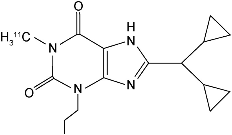
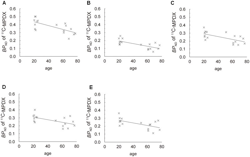
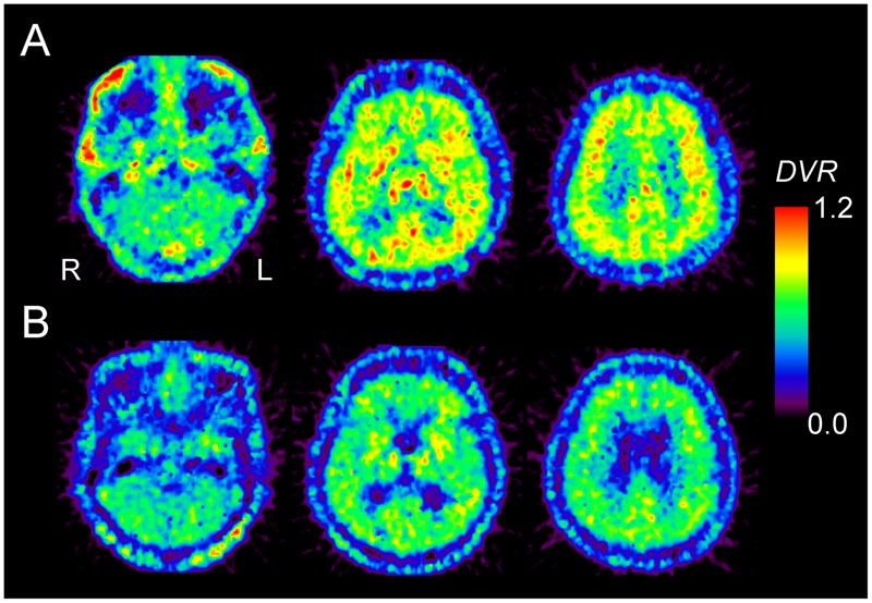
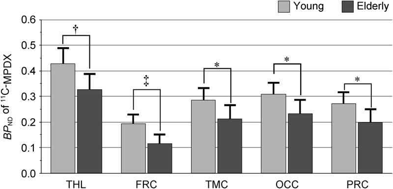
Adenosine A receptors (ARs) are widely distributed throughout the entire human brain, while adenosine A receptors (ARs) are present in dopamine-rich areas of the brain, such as the basal ganglia. A past study using autoradiography reported a reduced binding ability of AR in the striatum of old rats. We developed positron emission tomography (PET) ligands for mapping the adenosine receptors and we successfully visualized the ARs using 8-dicyclopropylmethyl-1-C-methyl-3-propylxanthine (C-MPDX). We previously reported that the density of ARs decreased with age in the human striatum, although we could not observe an age-related change in ARs. The aim of this study was to investigate the age-related change of the density of ARs in the thalamus and cerebral cortices of healthy participants using C-MPDX PET. We recruited eight young (22.0 ± 1.7 years) and nine elderly healthy male volunteers (65.7 ± 8.0 years). A dynamic series of decay-corrected PET scans was performed for 60 min starting with the injection of C-MPDX. We placed the circular regions of interest of 10 mm in diameter in C-MPDX PET images. The values for the binding potential () of C-MPDX in the thalamus, and frontal, temporal, occipital, and parietal cortices were calculated using a graphical analysis, wherein the reference region was the cerebellum. of C-MPDX was significantly lower in elderly participants than young participants in the thalamus, and frontal, temporal, occipital, and parietal cortices. In the human brain, we could observe the age-related decrease in the distribution of ARs.
腺苷 A 受体(ARs)广泛分布于整个人脑,而腺苷 A 受体(ARs)存在于大脑中富含多巴胺的区域,如基底神经节。过去一项使用放射自显影术的研究报告称,老年大鼠纹状体中 AR 的结合能力降低。我们开发了用于绘制腺苷受体图谱的正电子发射断层扫描(PET)配体,并使用 8 - 二环丙基甲基 - 1 - C - 甲基 - 3 - 丙基黄嘌呤(C - MPDX)成功可视化了 ARs。我们之前报道过,尽管未观察到 ARs 与年龄相关的变化,但人类纹状体中 ARs 的密度会随年龄下降。本研究的目的是使用 C - MPDX PET 研究健康参与者丘脑和大脑皮质中 ARs 密度与年龄相关的变化。我们招募了 8 名年轻(22.0 ± 1.7 岁)和 9 名老年健康男性志愿者(65.7 ± 8.0 岁)。从注射 C - MPDX 开始进行 60 分钟的动态衰减校正 PET 扫描。我们在 C - MPDX PET 图像中放置了直径为 10 毫米的圆形感兴趣区域。使用图形分析计算 C - MPDX 在丘脑以及额叶、颞叶、枕叶和顶叶皮质中的结合潜能(BP)值,其中参考区域为小脑。在丘脑以及额叶、颞叶、枕叶和顶叶皮质中,老年参与者的 C - MPDX 的 BP 显著低于年轻参与者。在人脑中,我们可以观察到 ARs 分布与年龄相关的下降。