Department of Neuroscience, University of Minnesota, Minneapolis, MN, USA.
Center for NeuroGenetics, Department of Molecular Genetics & Microbiology and Neurology, College of Medicine, Genetics Institute, University of Florida, Gainesville, FL, USA.
Neurobiol Dis. 2018 Apr;112:35-48. doi: 10.1016/j.nbd.2018.01.003. Epub 2018 Jan 10.
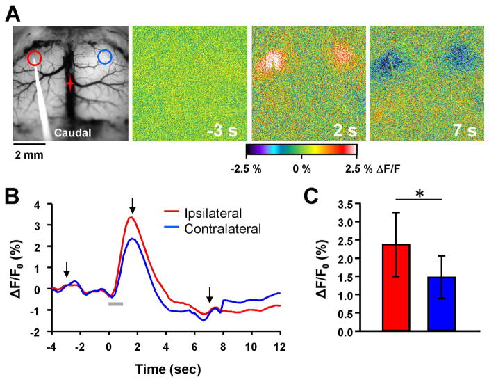
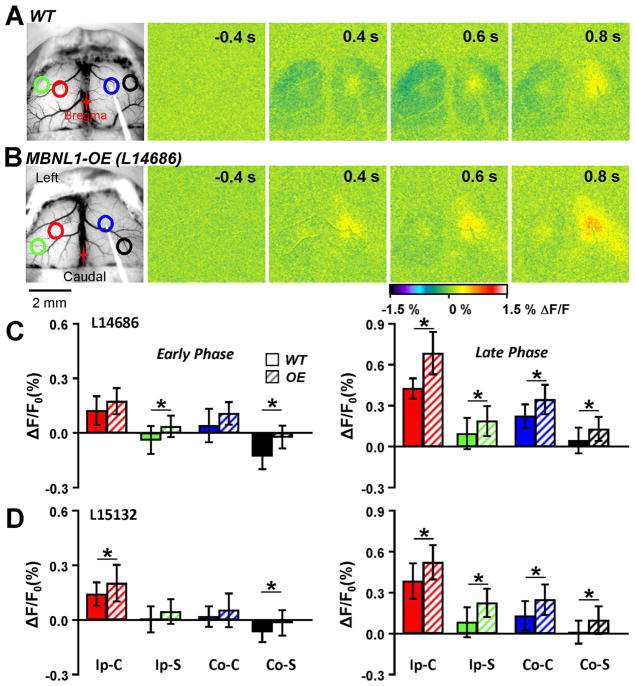
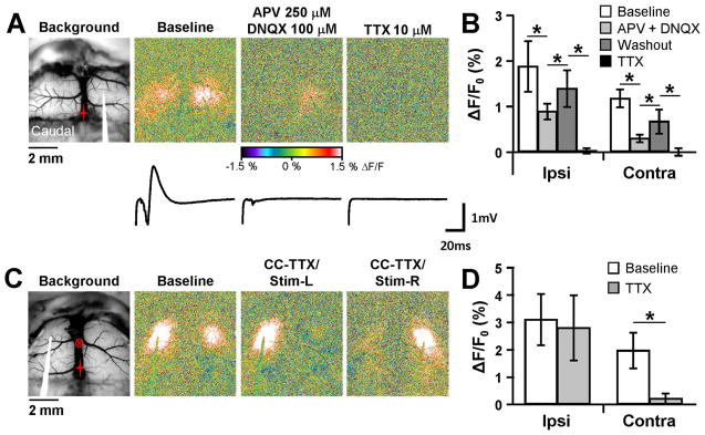
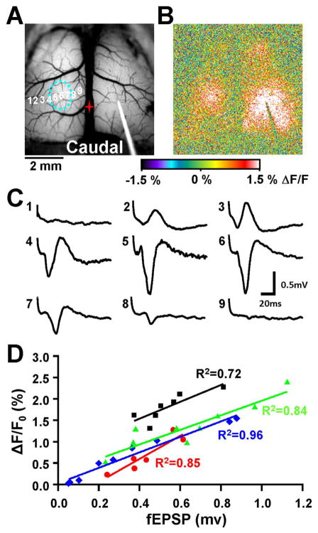
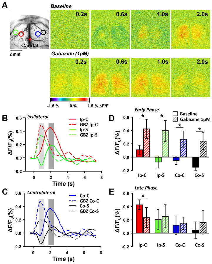
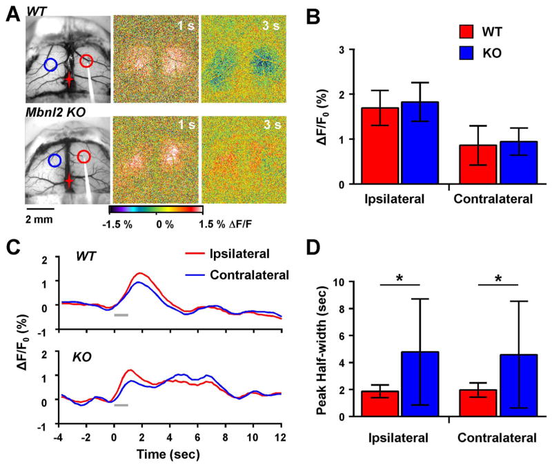
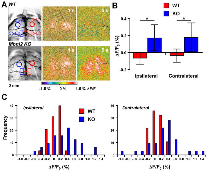
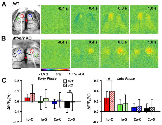
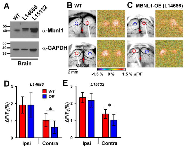
Myotonic dystrophy (DM) is a progressive, multisystem disorder affecting skeletal muscle, heart, and central nervous system. In both DM1 and DM2, microsatellite expansions of CUG and CCUG RNA repeats, respectively, accumulate and disrupt functions of alternative splicing factors, including muscleblind (MBNL) proteins. Grey matter loss and white matter changes, including the corpus callosum, likely underlie cognitive and executive function deficits in DM patients. However, little is known how cerebral cortical circuitry changes in DM. Here, flavoprotein optical imaging was used to assess local and contralateral responses to intracortical motor cortex stimulation in DM-related mouse models. In control mice, brief train stimulation generated ipsilateral and contralateral homotopic fluorescence increases, the latter mediated by the corpus callosum. Single pulse stimulation produced an excitatory response with an inhibitory-like surround response mediated by GABA receptors. In a mouse model of DM2 (Mbnl2 KO), we observed prolonged and increased responsiveness to train stimulation and loss of the inhibition from single pulse stimulation. Conversely, mice overexpressing human MBNL1 (MBNL1-OE) exhibited decreased contralateral response to train stimulation and reduction of inhibitory-like surround to single pulse stimulation. Therefore, altering levels of two key DM-associated splicing factors modifies functions of local cortical circuits and contralateral responses mediated through the corpus callosum.
肌强直性营养不良(DM)是一种进行性的多系统疾病,影响骨骼肌、心脏和中枢神经系统。在 DM1 和 DM2 中,分别有 CUG 和 CCUG RNA 重复微卫星的扩增,这些扩增会扰乱包括肌肉盲蛋白(MBNL)蛋白在内的可变剪接因子的功能。灰质丢失和白质改变,包括胼胝体,可能是 DM 患者认知和执行功能缺陷的基础。然而,对于 DM 中大脑皮质回路如何变化,我们知之甚少。在这里,使用黄素蛋白光学成像来评估 DM 相关小鼠模型中皮质内运动皮层刺激的局部和对侧反应。在对照小鼠中,短暂的训练刺激会产生同侧和对侧同源荧光增加,后者由胼胝体介导。单脉冲刺激产生兴奋性反应,周围抑制反应由 GABA 受体介导。在 DM2(Mbnl2 KO)的小鼠模型中,我们观察到对训练刺激的反应延长且增强,并且对单脉冲刺激的抑制作用丧失。相反,过表达人 MBNL1(MBNL1-OE)的小鼠对训练刺激的对侧反应降低,对单脉冲刺激的抑制性周围反应减少。因此,两种关键的 DM 相关剪接因子的水平改变会改变局部皮质回路和通过胼胝体介导的对侧反应的功能。