Serra Laura, Mancini Matteo, Silvestri Gabriella, Petrucci Antonio, Masciullo Marcella, Spanò Barbara, Torso Mario, Mastropasqua Chiara, Giacanelli Manlio, Caltagirone Carlo, Cercignani Mara, Meola Giovanni, Bozzali Marco
Neuroimaging Laboratory, IRCCS Santa Lucia Foundation, Via Ardeatina 306, 00179 Rome, Italy.
Neuroimaging Laboratory, IRCCS Santa Lucia Foundation, Via Ardeatina 306, 00179 Rome, Italy; Department of Engineering, "Roma Tre" University, Via Vito Volterra 62, 00154 Rome, Italy.
Neural Plast. 2016;2016:2696085. doi: 10.1155/2016/2696085. Epub 2016 May 25.
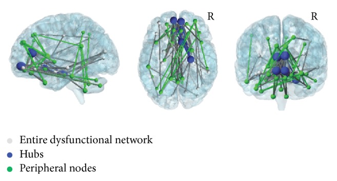
The adult form of myotonic dystrophy type 1 (DM1) presents with paradoxical inconsistencies between severity of brain damage, relative preservation of cognition, and failure in everyday life. This study, based on the assessment of brain connectivity and mechanisms of plasticity, aimed at reconciling these conflicting issues. Resting-state functional MRI and graph theoretical methods of analysis were used to assess brain topological features in a large cohort of patients with DM1. Patients, compared to controls, revealed reduced connectivity in a large frontoparietal network that correlated with their isolated impairment in visuospatial reasoning. Despite a global preservation of the topological properties, peculiar patterns of frontal disconnection and increased parietal-cerebellar connectivity were also identified in patients' brains. The balance between loss of connectivity and compensatory mechanisms in different brain networks might explain the paradoxical mismatch between structural brain damage and minimal cognitive deficits observed in these patients. This study provides a comprehensive assessment of brain abnormalities that fit well with both motor and nonmotor clinical features experienced by patients in their everyday life. The current findings suggest that measures of functional connectivity may offer the possibility of characterizing individual patients with the potential to become a clinical tool.
成人型1型强直性肌营养不良(DM1)表现出脑损伤严重程度、认知相对保留以及日常生活能力受损之间的矛盾不一致。本研究基于脑连接性和可塑性机制的评估,旨在调和这些相互矛盾的问题。采用静息态功能磁共振成像和图形理论分析方法,对一大群DM1患者的脑拓扑特征进行评估。与对照组相比,患者在一个大的额顶叶网络中显示出连接性降低,这与他们在视觉空间推理方面的孤立损伤相关。尽管拓扑属性整体得以保留,但在患者大脑中也发现了额叶断开连接的特殊模式以及顶叶 - 小脑连接性增加。不同脑网络中连接性丧失与代偿机制之间的平衡,可能解释了这些患者脑结构损伤与轻微认知缺陷之间的矛盾不匹配。本研究对脑异常进行了全面评估,这与患者在日常生活中经历的运动和非运动临床特征非常吻合。目前的研究结果表明,功能连接性测量可能为个体患者的特征描述提供可能性,有望成为一种临床工具。