Department of Radiology, the Fifth Affiliated Hospital, Sun Yat-sen University, Zhuhai 519000, China.
PCFM Lab of Ministry of Education, School of Materials Science and Engineering, Sun Yat-Sen University, Guangzhou 510275, China.
Int J Med Sci. 2018 Jan 1;15(2):129-141. doi: 10.7150/ijms.21610. eCollection 2018.
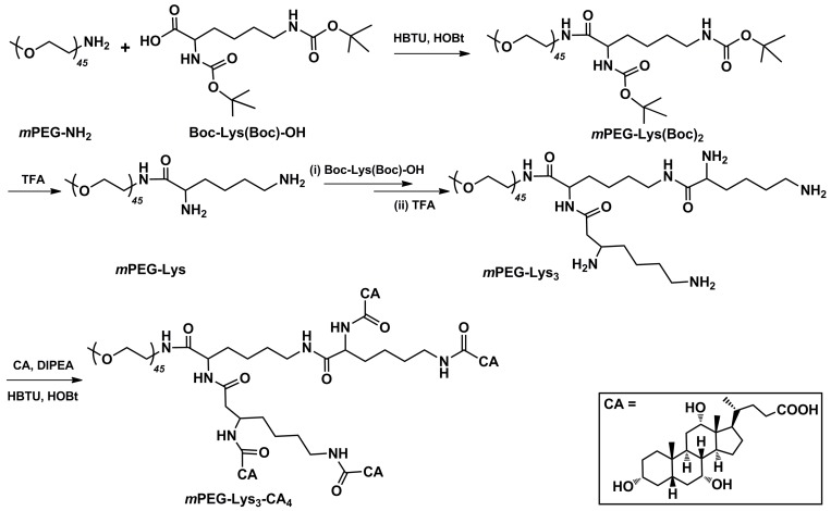
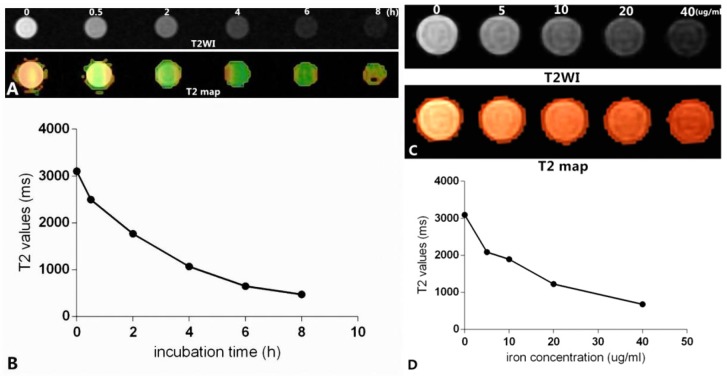
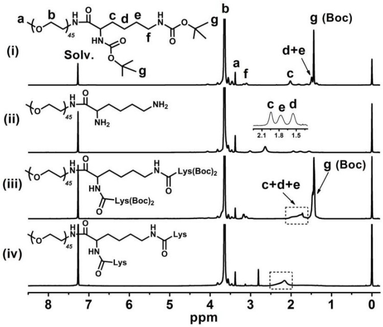
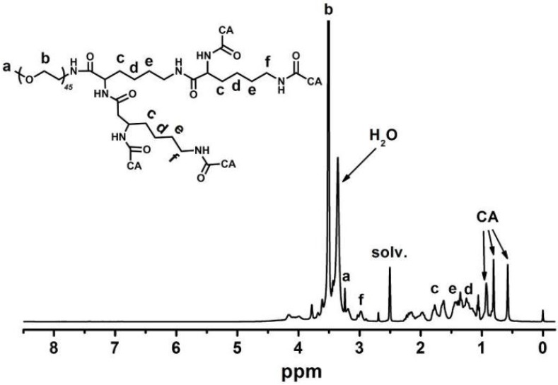
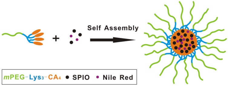
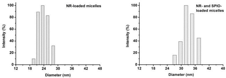
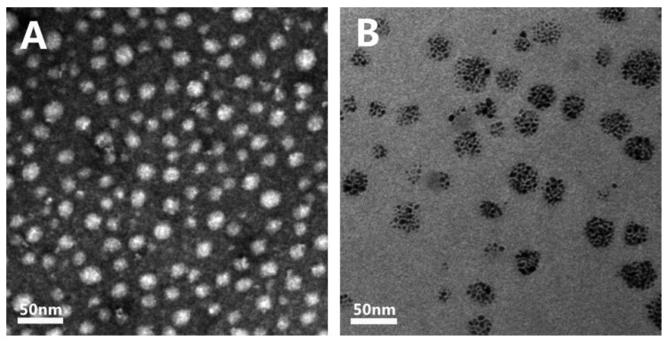
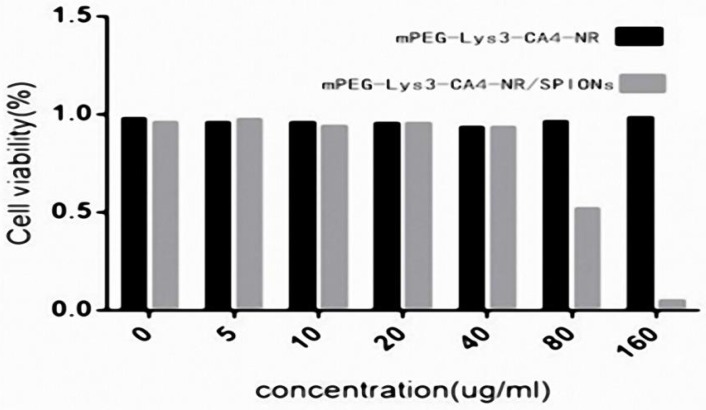
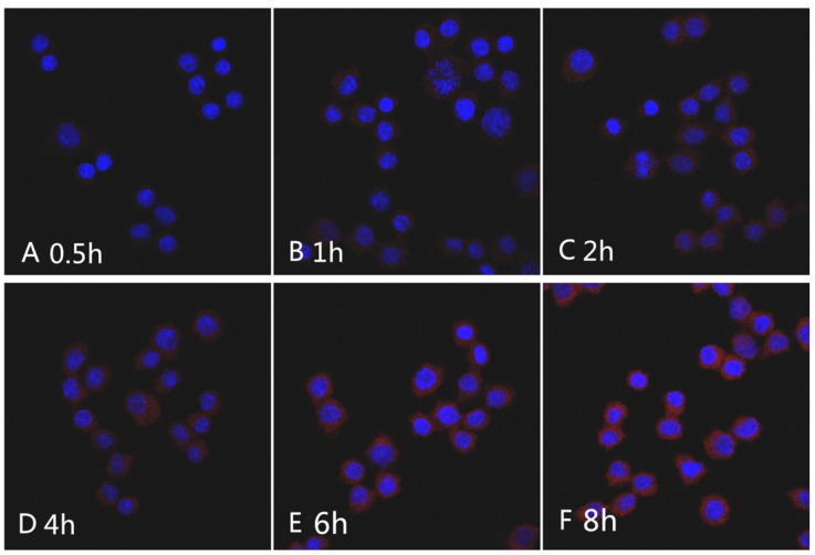
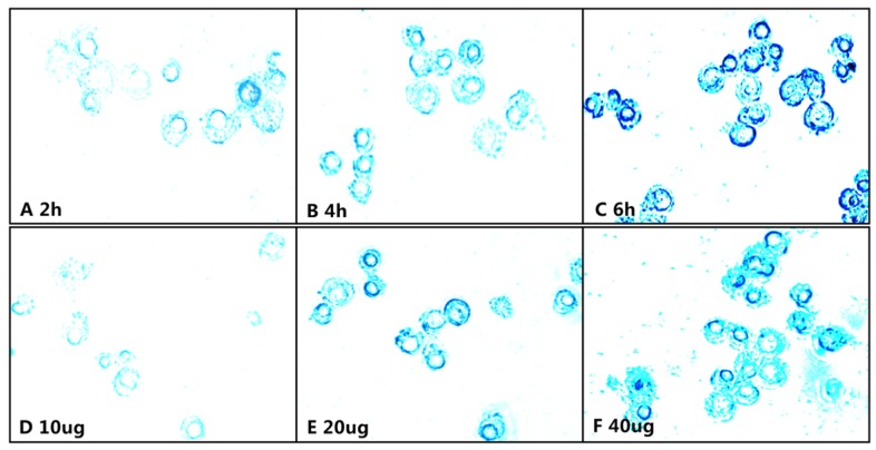
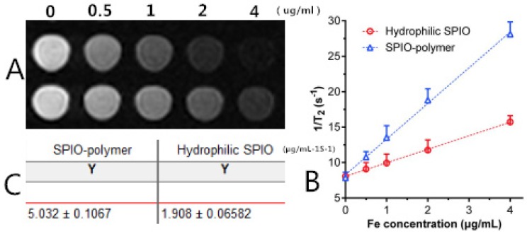
To establish small-sized superparamagnetic polymeric micelles for magnetic resonance and fluorescent dual-modal imaging, we investigated the feasibility of MR imaging (MRI) and macrophage-targeted in vitro. A new class of superparamagnetic iron oxide nanoparticles (SPIONs) and Nile red-co-loaded mPEG-Lys3-CA4-NR/SPION polymeric micelles was synthesized to label Raw264.7 cells. The physical characteristics of the polymeric micelles were assessed, the T2 relaxation rate was calculated, and the effect of labeling on the cell viability and cytotoxicity was also determined in vitro. In addition, further evaluation of the application potential of the micelles was conducted via in vitro MRI. The diameter of the mPEG-Lys3-CA4-NR/SPION polymeric micelles was 33.8 ± 5.8 nm on average. Compared with the hydrophilic SPIO, mPEG-Lys3-CA4-NR/SPION micelles increased transversely (r2), leading to a notably high r2 from 1.908 µg/mLS up to 5.032 µg/mLS, making the mPEG-Lys3-CA4-NR/SPION micelles a highly sensitive MRI T2 contrast agent, as further demonstrated by in vitro MRI. The results of Confocal Laser Scanning Microscopy (CLSM) and Prussian blue staining of Raw264.7 after incubation with micelle-containing medium indicated that the cellular uptake efficiency is high. We successfully synthesized dual-modal MR and fluorescence imaging mPEG-Lys3-CA4-NR/SPION polymeric micelles with an ultra-small size and high MRI sensitivity, which were effectively and quickly uptaken into Raw 264.7 cells. mPEG-Lys3-CA4-NR/SPION polymeric micelles might become a new MR lymphography contrast agent, with high effectiveness and high MRI sensitivity.
为了建立用于磁共振和荧光双模式成像的小尺寸超顺磁性聚合物胶束,我们研究了磁共振成像(MRI)和巨噬细胞靶向的可行性。合成了一种新型超顺磁性氧化铁纳米粒子(SPION)和尼罗红共载 mPEG-Lys3-CA4-NR/SPION 聚合物胶束来标记 Raw264.7 细胞。评估了聚合物胶束的物理特性,计算了 T2 弛豫率,并在体外测定了标记对细胞活力和细胞毒性的影响。此外,还通过体外 MRI 进一步评估了胶束的应用潜力。mPEG-Lys3-CA4-NR/SPION 聚合物胶束的平均直径为 33.8±5.8nm。与亲水性 SPIO 相比,mPEG-Lys3-CA4-NR/SPION 胶束增加了横向弛豫率(r2),导致 r2 从 1.908µg/mLS 显著增加至 5.032µg/mLS,使 mPEG-Lys3-CA4-NR/SPION 胶束成为一种高灵敏度的 MRI T2 对比剂,这也得到了体外 MRI 的进一步证实。载胶束培养基孵育后的 Raw264.7 的共聚焦激光扫描显微镜(CLSM)和普鲁士蓝染色结果表明,细胞摄取效率高。我们成功合成了具有超小尺寸和高 MRI 灵敏度的双模式磁共振和荧光成像 mPEG-Lys3-CA4-NR/SPION 聚合物胶束,能够有效快速地被 Raw264.7 细胞摄取。mPEG-Lys3-CA4-NR/SPION 聚合物胶束可能成为一种新的 MRI 淋巴管造影对比剂,具有高效性和高 MRI 灵敏度。