Yang Xue, Chung Jin-Yong, Rai Usha, Esumi Noriko
Wilmer Eye Institute, Johns Hopkins University School of Medicine, Baltimore, Maryland, United States of America.
PLoS One. 2018 Jan 16;13(1):e0191279. doi: 10.1371/journal.pone.0191279. eCollection 2018.
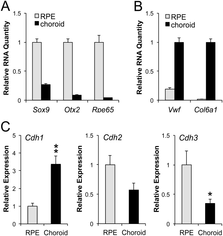
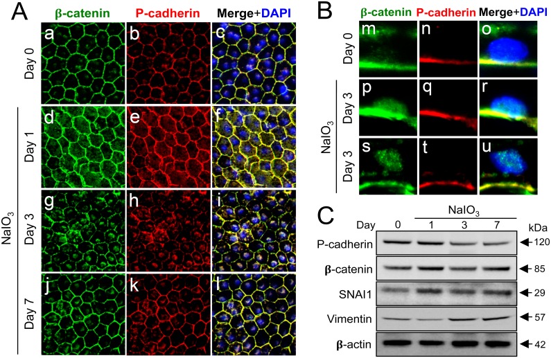
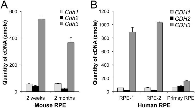
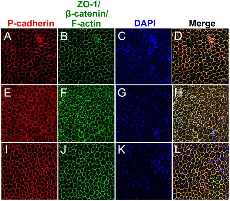
The retinal pigment epithelium (RPE) supports the health and function of retinal photoreceptors and is essential for normal vision. RPE cells are post-mitotic, terminally differentiated, and polarized epithelial cells. In pathological conditions, however, they lose their epithelial integrity, become dysfunctional, even dedifferentiate, and ultimately die. The integrity of epithelial cells is maintained, in part, by adherens junctions, which are composed of cadherin homodimers and p120-, β-, and α-catenins linking to actin filaments. While E-cadherin is the major cadherin for forming the epithelial phenotype in most epithelial cell types, it has been reported that cadherin expression in RPE cells is different from other epithelial cells based on results with cultured RPE cells. In this study, we revisited the expression of cadherins in the RPE to clarify their relative contribution by measuring the absolute quantity of cDNAs produced from mRNAs of three classical cadherins (E-, N-, and P-cadherins) in the RPE in vivo. We found that P-cadherin (CDH3) is highly dominant in both mouse and human RPE in situ. The degree of dominance of P-cadherin is surprisingly large, with mouse Cdh3 and human CDH3 accounting for 82-85% and 92-93% of the total of the three cadherin mRNAs, respectively. We confirmed the expression of P-cadherin protein at the cell-cell border of mouse RPE in situ by immunofluorescence. Furthermore, we found that oxidative stress induces dissociation of P-cadherin and β-catenin from the cell membrane and subsequent translocation of β-catenin into the nucleus, resulting in activation of the canonical Wnt/β-catenin pathway. This is the first report of absolute comparison of the expression of three cadherins in the RPE, and the results suggest that the physiological role of P-cadherin in the RPE needs to be reevaluated.
视网膜色素上皮(RPE)维持视网膜光感受器的健康与功能,对正常视觉至关重要。RPE细胞是终末分化的、有丝分裂后且极化的上皮细胞。然而,在病理状态下,它们会丧失上皮完整性,功能失调,甚至去分化,最终死亡。上皮细胞的完整性部分由黏附连接维持,黏附连接由钙黏蛋白同型二聚体以及连接肌动蛋白丝的p120、β和α连环蛋白组成。虽然E-钙黏蛋白是大多数上皮细胞类型中形成上皮表型的主要钙黏蛋白,但根据培养的RPE细胞的结果,有报道称RPE细胞中钙黏蛋白的表达与其他上皮细胞不同。在本研究中,我们通过测量体内RPE中三种经典钙黏蛋白(E-、N-和P-钙黏蛋白)mRNA产生的cDNA的绝对量,重新审视了RPE中钙黏蛋白的表达,以阐明它们的相对贡献。我们发现,P-钙黏蛋白(CDH3)在小鼠和人类RPE原位均高度占主导地位。P-钙黏蛋白的主导程度惊人地高,小鼠Cdh3和人类CDH3分别占三种钙黏蛋白mRNA总量的82 - 85%和92 - 93%。我们通过免疫荧光证实了小鼠RPE原位细胞 - 细胞边界处P-钙黏蛋白的表达。此外,我们发现氧化应激诱导P-钙黏蛋白和β-连环蛋白从细胞膜解离,随后β-连环蛋白易位至细胞核,导致经典Wnt/β-连环蛋白通路激活。这是首次对RPE中三种钙黏蛋白的表达进行绝对比较的报告,结果表明需要重新评估P-钙黏蛋白在RPE中的生理作用。