• 文献检索
  • 文档翻译
  • 深度研究
  • 学术资讯
  • Suppr Zotero 插件Zotero 插件
  • 邀请有礼
  • 套餐&价格
  • 历史记录
应用&插件
Suppr Zotero 插件Zotero 插件浏览器插件Mac 客户端Windows 客户端微信小程序
定价
高级版会员购买积分包购买API积分包
服务
文献检索文档翻译深度研究API 文档MCP 服务
关于我们
关于 Suppr公司介绍联系我们用户协议隐私条款
关注我们

Suppr 超能文献

核心技术专利:CN118964589B侵权必究
粤ICP备2023148730 号-1Suppr @ 2026

文献检索

告别复杂PubMed语法,用中文像聊天一样搜索,搜遍4000万医学文献。AI智能推荐,让科研检索更轻松。

立即免费搜索

文件翻译

保留排版,准确专业,支持PDF/Word/PPT等文件格式,支持 12+语言互译。

免费翻译文档

深度研究

AI帮你快速写综述,25分钟生成高质量综述,智能提取关键信息,辅助科研写作。

立即免费体验

血管生成素样蛋白 4 在去甲肾上腺素诱导的心肌细胞肥大中的作用。

The role of angiopoietin-like protein 4 in phenylephrine-induced cardiomyocyte hypertrophy.

机构信息

Department of Cardiology, the First Affiliated Hospital of Sun Yat-Sen University, Guangzhou, Guangdong, China, 510080.

Key Laboratory on Assisted Circulation, Ministry of Health, Guangzhou, Guangdong, China, 510080.

出版信息

Biosci Rep. 2019 Jul 29;39(7). doi: 10.1042/BSR20171358. Print 2019 Jul 31.

DOI:10.1042/BSR20171358
PMID:29339422
原文链接:https://pmc.ncbi.nlm.nih.gov/articles/PMC6663991/
Abstract

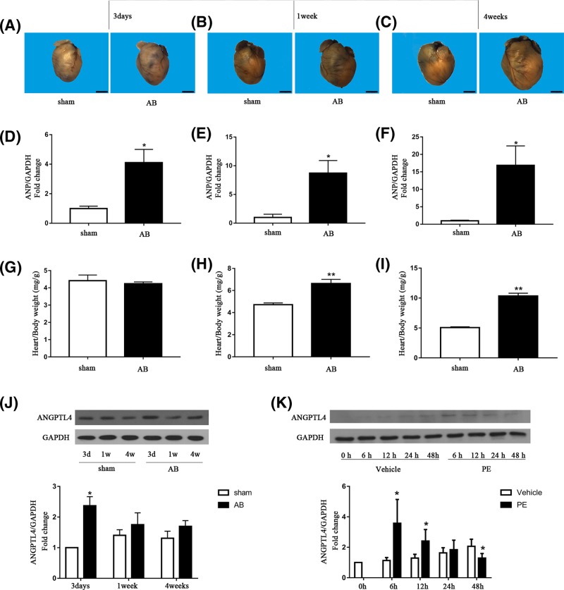
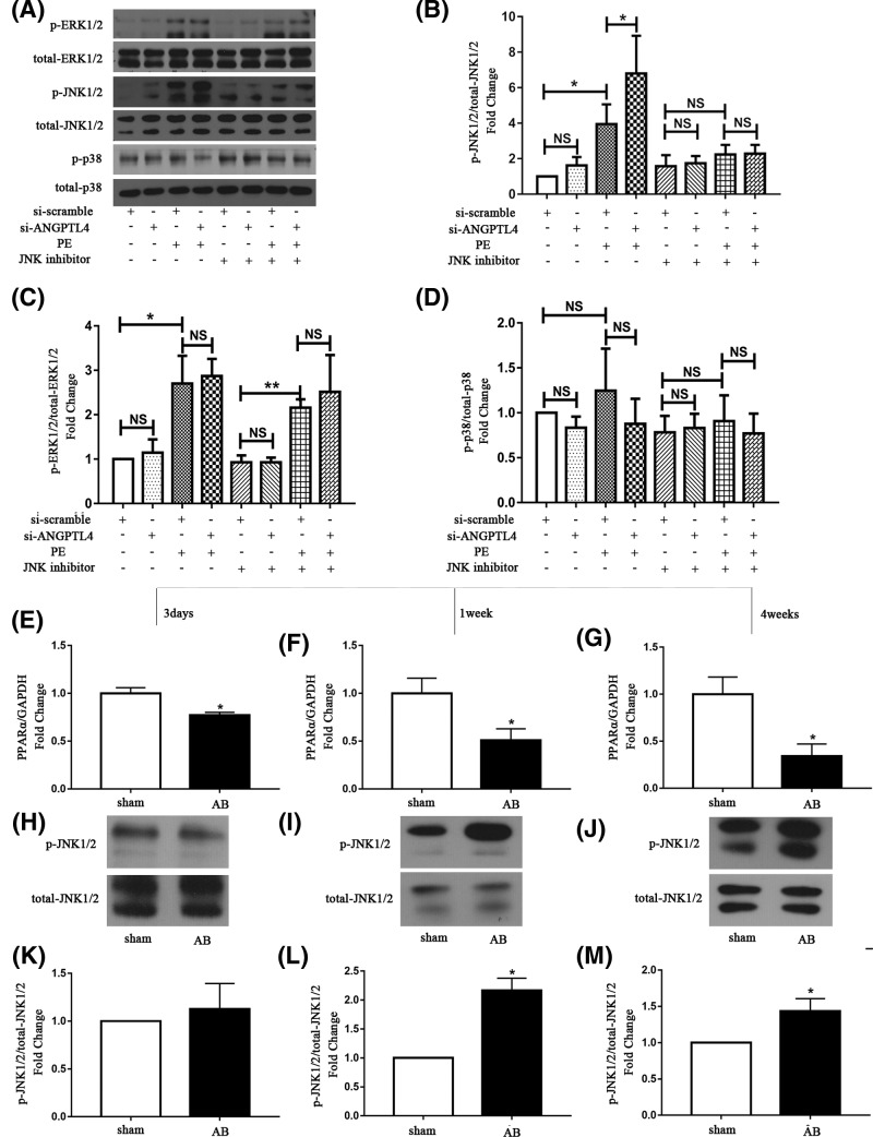
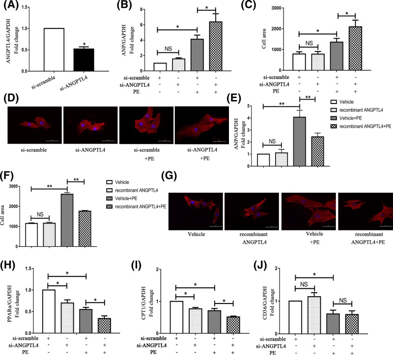
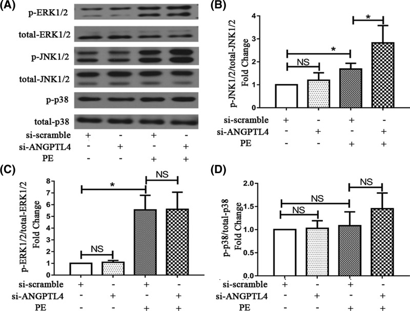
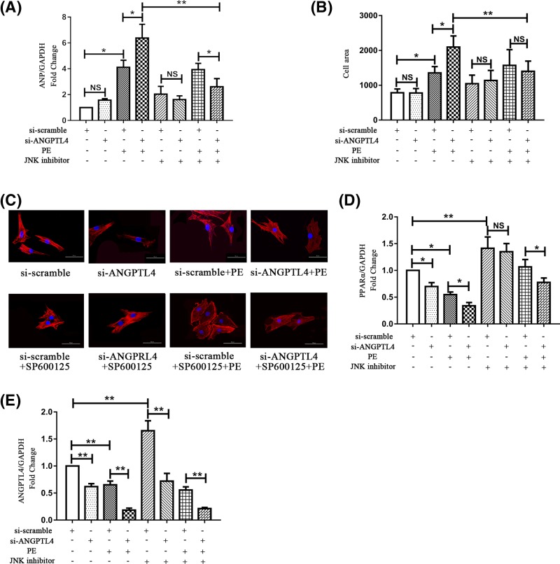
Angiopoietin-like protein 4 (ANGPTL4) is a multifunctional secreted protein that can be induced by fasting, hypoxia and glucocorticoids. ANGPTL4 has been associated with a variety of diseases; however, the role of ANGPTL4 in cardiac hypertrophy remains poorly understood. In our study, we aimed to explore the effect of ANGPTL4 on phenylephrine-induced cardiomyocyte hypertrophy. Our results showed that knockdown of ANGPTL4 expression significantly exacerbated cardiomyocyte hypertrophy, as demonstrated by increased hypertrophic marker expression, including ANP and cell surface area. Moreover, significantly reduced fatty acid oxidation, as featured by decreased CPT-1 levels, was observed in hypertrophic cardiomyocytes following ANGPTL4 down-regulation. Furthermore, knockdown of ANGPLT4 led to down-regulated expression of peroxisome proliferator-activated receptor α (PPARα), which is the key regulator of cardiac fatty acid oxidation. In addition, ANGPTL4 silencing promoted the activation of JNK1/2, and JNK1/2 signaling blockade could restore the level of PPARα and significantly ameliorate the ANGPTL4 knockdown-induced cardiomyocyte hypertrophy. Therefore, our study demonstrated that ANGPTL4 regulates PPARα through JNK1/2 signaling and is required for the inhibition of cardiomyocyte hypertrophy.

摘要

血管生成素样蛋白 4(ANGPTL4)是一种多功能分泌蛋白,可被禁食、缺氧和糖皮质激素诱导。ANGPTL4 与多种疾病有关;然而,ANGPTL4 在心肌肥厚中的作用仍知之甚少。在我们的研究中,我们旨在探讨 ANGPTL4 对苯肾上腺素诱导的心肌细胞肥大的影响。我们的结果表明,ANGPTL4 表达的敲低显著加剧了心肌细胞肥大,表现为肥大标志物的表达增加,包括 ANP 和细胞表面积。此外,在 ANGPTL4 下调后,肥大心肌细胞中的脂肪酸氧化明显减少,表现为 CPT-1 水平降低。此外,ANGPLT4 的敲低导致过氧化物酶体增殖物激活受体 α(PPARα)的表达下调,PPARα 是心脏脂肪酸氧化的关键调节因子。此外,ANGPTL4 沉默促进了 JNK1/2 的激活,而 JNK1/2 信号通路的阻断可以恢复 PPARα 的水平,并显著改善 ANGPTL4 敲低诱导的心肌细胞肥大。因此,我们的研究表明,ANGPTL4 通过 JNK1/2 信号通路调节 PPARα,是抑制心肌细胞肥大所必需的。

https://cdn.ncbi.nlm.nih.gov/pmc/blobs/87ce/6663991/870365d50720/bsr-39-bsr20171358-g5.jpg
https://cdn.ncbi.nlm.nih.gov/pmc/blobs/87ce/6663991/62027dcd2a92/bsr-39-bsr20171358-g1.jpg
https://cdn.ncbi.nlm.nih.gov/pmc/blobs/87ce/6663991/81704a19246f/bsr-39-bsr20171358-g2.jpg
https://cdn.ncbi.nlm.nih.gov/pmc/blobs/87ce/6663991/b85c9402e786/bsr-39-bsr20171358-g3.jpg
https://cdn.ncbi.nlm.nih.gov/pmc/blobs/87ce/6663991/5b7f7b2c23dc/bsr-39-bsr20171358-g4.jpg
https://cdn.ncbi.nlm.nih.gov/pmc/blobs/87ce/6663991/870365d50720/bsr-39-bsr20171358-g5.jpg

相似文献

1
The role of angiopoietin-like protein 4 in phenylephrine-induced cardiomyocyte hypertrophy.血管生成素样蛋白 4 在去甲肾上腺素诱导的心肌细胞肥大中的作用。
Biosci Rep. 2019 Jul 29;39(7). doi: 10.1042/BSR20171358. Print 2019 Jul 31.
2
Effects of ERK1/2/PPARα/SCAD signal pathways on cardiomyocyte hypertrophy induced by insulin-like growth factor 1 and phenylephrine.胰岛素样生长因子 1 和苯肾上腺素诱导的心肌细胞肥大的 ERK1/2/PPARα/SCAD 信号通路的作用。
Life Sci. 2015 Mar 1;124:41-9. doi: 10.1016/j.lfs.2015.01.015. Epub 2015 Jan 28.
3
Sestrin 2 attenuates neonatal rat cardiomyocyte hypertrophy induced by phenylephrine via inhibiting ERK1/2.Sesnrin 2 通过抑制 ERK1/2 减轻苯肾上腺素诱导的新生大鼠心肌细胞肥大。
Mol Cell Biochem. 2017 Sep;433(1-2):113-123. doi: 10.1007/s11010-017-3020-2. Epub 2017 May 11.
4
Hypoxia-Induced Mitogenic Factor Promotes Cardiac Hypertrophy via Calcium-Dependent and Hypoxia-Inducible Factor-1α Mechanisms.缺氧诱导的有丝分裂因子通过钙依赖性和缺氧诱导因子-1α 机制促进心肌肥大。
Hypertension. 2018 Aug;72(2):331-342. doi: 10.1161/HYPERTENSIONAHA.118.10845. Epub 2018 Jun 11.
5
Adenosine monophosphate-activated protein kinase inhibits cardiac hypertrophy through reactivating peroxisome proliferator-activated receptor-alpha signaling pathway.单磷酸腺苷激活的蛋白激酶通过重新激活过氧化物酶体增殖物激活受体α信号通路来抑制心肌肥大。
Eur J Pharmacol. 2009 Oct 12;620(1-3):63-70. doi: 10.1016/j.ejphar.2009.08.024. Epub 2009 Aug 20.
6
Downregulation of adipose triglyceride lipase promotes cardiomyocyte hypertrophy by triggering the accumulation of ceramides.脂肪甘油三酯脂肪酶的下调通过引发神经酰胺的积累促进心肌细胞肥大。
Arch Biochem Biophys. 2015 Jan 1;565:76-88. doi: 10.1016/j.abb.2014.11.009. Epub 2014 Nov 28.
7
Changes in short-chain acyl-coA dehydrogenase during rat cardiac development and stress.大鼠心脏发育和应激过程中短链酰基辅酶A脱氢酶的变化
J Cell Mol Med. 2015 Jul;19(7):1672-88. doi: 10.1111/jcmm.12541. Epub 2015 Mar 8.
8
Sirt1 acts in association with PPARα to protect the heart from hypertrophy, metabolic dysregulation, and inflammation.Sirt1 与 PPARα 协同作用,保护心脏免受肥大、代谢失调和炎症的影响。
Cardiovasc Res. 2011 May 1;90(2):276-84. doi: 10.1093/cvr/cvq376. Epub 2010 Nov 29.
9
Targeting Peroxisome Proliferator Activated Receptor α (PPAR α) for the Prevention of Mitochondrial Impairment and Hypertrophy in Cardiomyocytes.靶向过氧化物酶体增殖物激活受体α(PPARα)预防心肌细胞线粒体损伤和肥大
Cell Physiol Biochem. 2018;49(1):245-259. doi: 10.1159/000492875. Epub 2018 Aug 23.
10
PEX5 prevents cardiomyocyte hypertrophy via suppressing the redox-sensitive signaling pathways MAPKs and STAT3.PEX5 通过抑制氧化还原敏感的信号通路 MAPKs 和 STAT3 来防止心肌细胞肥大。
Eur J Pharmacol. 2021 Sep 5;906:174283. doi: 10.1016/j.ejphar.2021.174283. Epub 2021 Jun 24.

引用本文的文献

1
ANGPTL4 suppresses the profibrogenic functions of atrial fibroblasts induced by angiotensin II by up-regulating PPARγ.血管生成素样蛋白4通过上调过氧化物酶体增殖物激活受体γ来抑制血管紧张素II诱导的心房成纤维细胞的促纤维化功能。
Iran J Basic Med Sci. 2023;26(5):587-593. doi: 10.22038/IJBMS.2023.69196.15077.
2
ANGPTL4 Attenuates Ang II-Induced Atrial Fibrillation and Fibrosis in Mice via PPAR Pathway.血管生成素样蛋白4通过PPAR途径减轻血管紧张素II诱导的小鼠心房颤动和纤维化。
Cardiol Res Pract. 2021 Aug 9;2021:9935310. doi: 10.1155/2021/9935310. eCollection 2021.
3
Relationship between serum angiopoietin-like 4 levels and recurrence of atrial fibrillation.

本文引用的文献

1
Sestrin 2 attenuates neonatal rat cardiomyocyte hypertrophy induced by phenylephrine via inhibiting ERK1/2.Sesnrin 2 通过抑制 ERK1/2 减轻苯肾上腺素诱导的新生大鼠心肌细胞肥大。
Mol Cell Biochem. 2017 Sep;433(1-2):113-123. doi: 10.1007/s11010-017-3020-2. Epub 2017 May 11.
2
Sestrin 1 ameliorates cardiac hypertrophy via autophagy activation.硒蛋白1通过激活自噬改善心肌肥大。
J Cell Mol Med. 2017 Jun;21(6):1193-1205. doi: 10.1111/jcmm.13052. Epub 2017 Feb 9.
3
The Failing Heart Relies on Ketone Bodies as a Fuel.衰竭心脏依赖酮体作为燃料。
血清血管生成素样蛋白 4 水平与心房颤动复发的关系。
J Int Med Res. 2021 Feb;49(2):300060520988393. doi: 10.1177/0300060520988393.
4
Maf1 ameliorates cardiac hypertrophy by inhibiting RNA polymerase III through ERK1/2.Maf1 通过 ERK1/2 抑制 RNA 聚合酶 III 来改善心肌肥厚。
Theranostics. 2019 Sep 25;9(24):7268-7281. doi: 10.7150/thno.33006. eCollection 2019.
5
Stimulation of Na/K-ATPase with an Antibody against Its 4 Extracellular Region Attenuates Angiotensin II-Induced H9c2 Cardiomyocyte Hypertrophy via an AMPK/SIRT3/PPAR Signaling Pathway.用针对其 4 个细胞外区的抗体刺激 Na/K-ATPase 可通过 AMPK/SIRT3/PPAR 信号通路减轻血管紧张素 II 诱导的 H9c2 心肌细胞肥大。
Oxid Med Cell Longev. 2019 Sep 15;2019:4616034. doi: 10.1155/2019/4616034. eCollection 2019.
Circulation. 2016 Feb 23;133(8):698-705. doi: 10.1161/CIRCULATIONAHA.115.017355. Epub 2016 Jan 27.
4
Evidence for Intramyocardial Disruption of Lipid Metabolism and Increased Myocardial Ketone Utilization in Advanced Human Heart Failure.晚期人类心力衰竭中心肌内脂质代谢紊乱及心肌酮利用增加的证据。
Circulation. 2016 Feb 23;133(8):706-16. doi: 10.1161/CIRCULATIONAHA.115.017545. Epub 2016 Jan 27.
5
REDD1 attenuates cardiac hypertrophy via enhancing autophagy.REDD1通过增强自噬来减轻心脏肥大。
Biochem Biophys Res Commun. 2014 Nov 7;454(1):215-20. doi: 10.1016/j.bbrc.2014.10.079. Epub 2014 Oct 22.
6
Revisiting PPARγ as a target for the treatment of metabolic disorders.重新审视 PPARγ 作为治疗代谢紊乱的靶点。
BMB Rep. 2014 Nov;47(11):599-608. doi: 10.5483/bmbrep.2014.47.11.174.
7
The PPARα-FGF21 hormone axis contributes to metabolic regulation by the hepatic JNK signaling pathway.过氧化物酶体增殖物激活受体α-成纤维细胞生长因子21激素轴通过肝脏c-Jun氨基末端激酶信号通路参与代谢调节。
Cell Metab. 2014 Sep 2;20(3):512-25. doi: 10.1016/j.cmet.2014.06.010. Epub 2014 Jul 17.
8
AMPK inhibits cardiac hypertrophy by promoting autophagy via mTORC1.AMPK 通过抑制 mTORC1 促进自噬来抑制心肌肥厚。
Arch Biochem Biophys. 2014 Sep 15;558:79-86. doi: 10.1016/j.abb.2014.06.023. Epub 2014 Jul 4.
9
Cardiac metabolism and its interactions with contraction, growth, and survival of cardiomyocytes.心肌代谢及其与心肌细胞收缩、生长和存活的相互作用。
Circ Res. 2013 Aug 16;113(5):603-16. doi: 10.1161/CIRCRESAHA.113.302095.
10
Effect of Berberine on PPAR α /NO Activation in High Glucose- and Insulin-Induced Cardiomyocyte Hypertrophy.小檗碱对高糖和胰岛素诱导的心肌细胞肥大中 PPARα/NO 激活的影响。
Evid Based Complement Alternat Med. 2013;2013:285489. doi: 10.1155/2013/285489. Epub 2013 Mar 20.