Farokhian Farnaz, Yang Chunlan, Beheshti Iman, Matsuda Hiroshi, Wu Shuicai
1College of Life Science and Bioengineering, Beijing University of Technology, Beijing, 100022, China.
2Integrative Brain Imaging Center, National Center of Neurology and Psychiatry, Kodaira, Tokyo Japan.
Aging Dis. 2017 Dec 1;8(6):899-909. doi: 10.14336/AD.2017.0502. eCollection 2017 Dec.
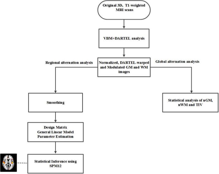
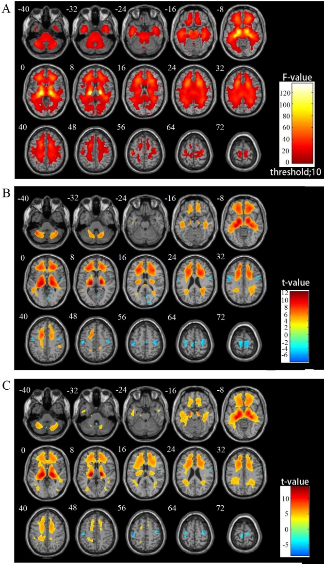
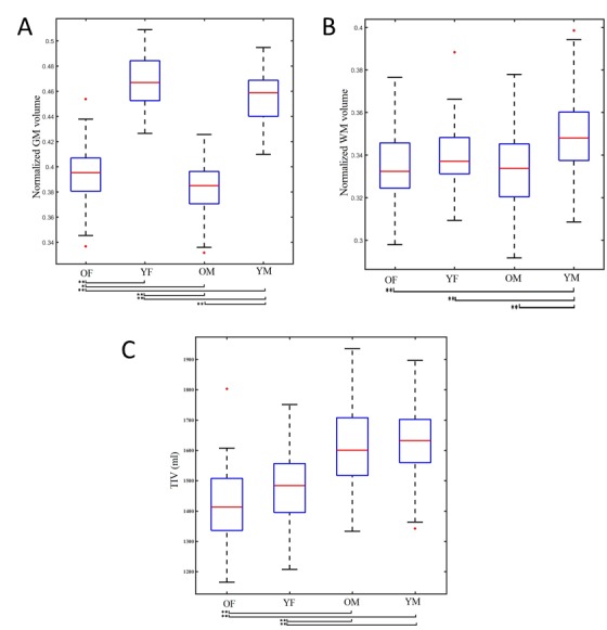
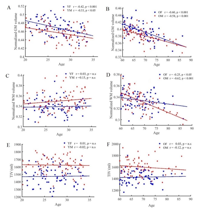
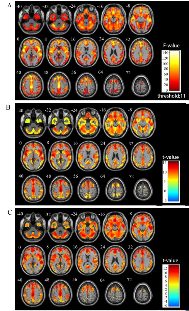
Normal aging is associated with both structural changes in many brain regions and functional declines in several cognitive domains with advancing age. Advanced neuroimaging techniques enable explorative analyses of structural alterations that can be used as assessments of such age-related changes. Here we used voxel-based morphometry (VBM) to investigate regional and global brain volume differences among four groups of healthy adults from the IXI Dataset: older females (OF, mean age 68.35 yrs; n=69), older males (OM, 68.43 yrs; n=66), young females (YF, 27.09 yrs; n=71), and young males (YM, 27.91 yrs; n=71), using 3D T1-weighted MRI data. At the global level, we investigated the influence of age and gender on brain volumes using a two-way analysis of variance. With respect to gender, we used the Pearson correlation to investigate global brain volume alterations due to age in the older and young groups. At the regional level, we used a flexible factorial statistical test to compare the means of gray matter (GM) and white matter (WM) volume alterations among the four groups. We observed different patterns in both the global and regional GM and WM alterations in the young and older groups with respect to gender. At the global level, we observed significant influences of age and gender on global brain volumes. At the regional level, the older subjects showed a widespread reduction in GM volume in regions of the frontal, insular, and cingulate cortices compared to the young subjects in both genders. Compared to the young subjects, the older subjects showed a widespread WM decline prominently in the thalamic radiations, in addition to increased WM in pericentral and occipital areas. Knowledge of these observed brain volume differences and changes may contribute to the elucidation of mechanisms underlying aging as well as age-related brain atrophy and disease.
正常衰老与许多脑区的结构变化以及随着年龄增长多个认知领域的功能衰退相关。先进的神经成像技术能够对结构改变进行探索性分析,这些结构改变可用于评估此类与年龄相关的变化。在这里,我们使用基于体素的形态计量学(VBM),利用三维T1加权磁共振成像(MRI)数据,研究了来自IXI数据集的四组健康成年人的区域和全脑体积差异:老年女性(OF,平均年龄68.35岁;n = 69)、老年男性(OM,68.43岁;n = 66)、年轻女性(YF,27.09岁;n = 71)和年轻男性(YM,27.91岁;n = 71)。在全脑水平上,我们使用双向方差分析研究年龄和性别对脑体积的影响。关于性别,我们使用Pearson相关性来研究老年组和年轻组中由于年龄导致的全脑体积变化。在区域水平上,我们使用灵活的析因统计检验来比较四组之间灰质(GM)和白质(WM)体积变化的均值。我们观察到,在年轻组和老年组中,GM和WM的全脑及区域变化在性别方面呈现出不同模式。在全脑水平上,我们观察到年龄和性别对全脑体积有显著影响。在区域水平上,与两个性别的年轻受试者相比,老年受试者额叶、岛叶和扣带回皮质区域的GM体积普遍减少。与年轻受试者相比,老年受试者除了中央周围和枕叶区域的WM增加外,丘脑辐射区域的WM也显著普遍下降。了解这些观察到的脑体积差异和变化可能有助于阐明衰老以及与年龄相关的脑萎缩和疾病的潜在机制。