Hajihassani Neda, Roohi Neda, Madadi Karim, Bakhshi Mahin, Tofangchiha Maryam
Department of Endodontics, Dental School, Qazvin University of Medical Sciences, Qazvin, Iran.
Private Practice, First Floor, No. 1, 7th Ave, Shohada Blvd, P.O. Box 3175745116, Fardis, Kara, Alborz Province, Iran.
Scientifica (Cairo). 2017;2017:1504341. doi: 10.1155/2017/1504341. Epub 2017 Nov 16.
Successful dental root canal treatments require a complete knowledge of dental anatomy and root canal morphology.
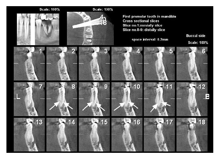
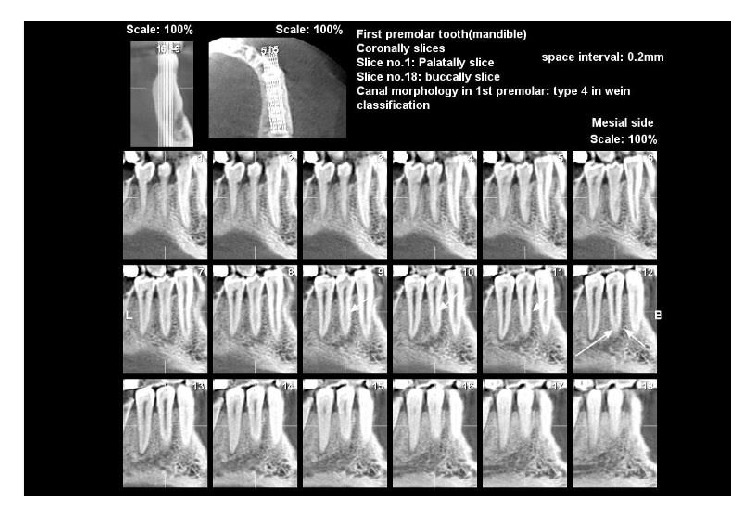
One hundred and forty-five cone beam computed tomography (CBCT) images were used to assess the anatomy and morphology of mandibular premolars based on Vertucci's classifications in a defined group of dental patients in Iran. The number of roots and root canals, root canal morphology, root and canal shape (curvature), existence of C-shaped canal, and influence of sex on each of these were evaluated. A chi-squared test was used for statistical analysis.
The mandibular first and second premolars had a single root in 95.97% and 100% cases, respectively. In the mandibular first premolars, 62.2% were of type I, 0.8% type II, 10.9% type III, 0.8% type IV, 20.3% type V, 4.2% type VI, and 0.8% type VII; in the second premolars, 78% of canals were of type I, 3% type II, 11% type III, 7% type V, and 1% type VI. C-shaped canals did not exist in either of the premolars. The most prevalent root and canal shape was straight. The most prevalent root curvature was a distal curvature in both premolars (71.4% and 74% of first and second premolars, resp.). The most prevalent canal curvature was lingual and buccal for the first premolars (7.6% each) and distal for the second premolars (11%). No significant difference was found between men and women in nearly all of the above ( > 0.05).
The results suggest that there is a need to conduct further evaluations on finding root and canal variations among more populations to gain better knowledge prior to root canal treatment.
成功的牙髓根管治疗需要全面了解牙齿解剖结构和根管形态。
在伊朗一组特定的牙科患者中,使用145张锥形束计算机断层扫描(CBCT)图像,根据韦尔图奇分类法评估下颌前磨牙的解剖结构和形态。评估了牙根和根管的数量、根管形态、牙根和根管形状(弯曲度)、C形根管的存在情况以及性别对上述各项的影响。采用卡方检验进行统计分析。
下颌第一前磨牙和第二前磨牙分别有95.97%和100%的病例为单根。在下颌第一前磨牙中,I型占62.2%,II型占0.8%,III型占10.9%,IV型占0.8%,V型占20.3%,VI型占4.2%,VII型占0.8%;在第二前磨牙中,78%的根管为I型,3%为II型,11%为III型,7%为V型,1%为VI型。两种前磨牙均未发现C形根管。最常见的牙根和根管形状是直的。两种前磨牙中最常见的牙根弯曲是远中弯曲(第一前磨牙和第二前磨牙分别为71.4%和74%)。第一前磨牙最常见的根管弯曲是舌侧和颊侧(各占7.6%),第二前磨牙是远中弯曲(占11%)。在几乎所有上述方面,男性和女性之间均未发现显著差异(>0.05)。
结果表明,在进行根管治疗之前需要对更多人群的牙根和根管变异进行进一步评估,以获取更全面的了解。