Division of Pharmacology and Pharmacotherapy, University of Helsinki, Viikinkaari 5E, P.O. Box 56, 00014, Helsinki, Finland.
Institute of Biotechnology, Research Program in Developmental Biology, University of Helsinki, Viikinkaari 5D, P.O. Box 56, 00014, Helsinki, Finland.
Mol Neurobiol. 2018 Aug;55(8):6755-6768. doi: 10.1007/s12035-018-0872-8. Epub 2018 Jan 18.
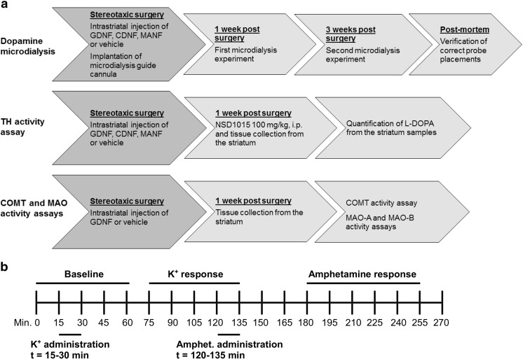
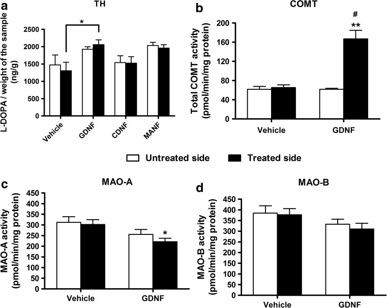
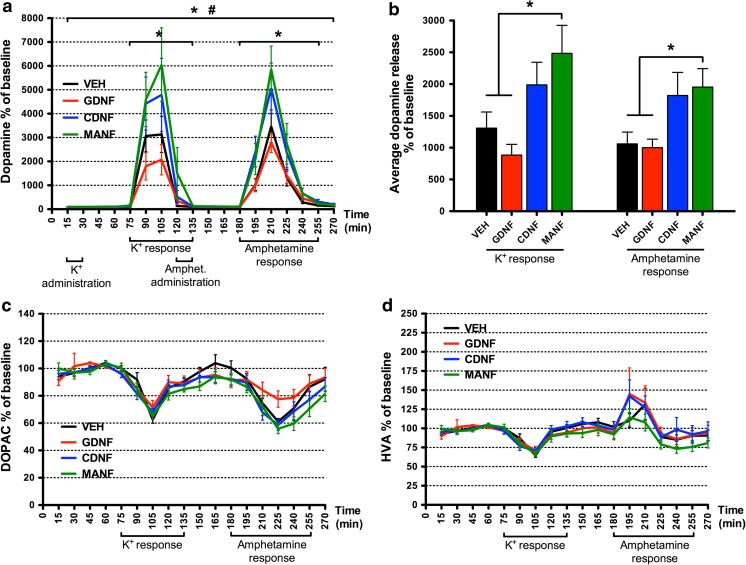
Neurotrophic factors (NTFs) hold potential as disease-modifying therapies for neurodegenerative disorders like Parkinson's disease. Glial cell line-derived neurotrophic factor (GDNF), cerebral dopamine neurotrophic factor (CDNF), and mesencephalic astrocyte-derived neurotrophic factor (MANF) have shown neuroprotective and restorative effects on nigral dopaminergic neurons in various animal models of Parkinson's disease. To date, however, their effects on brain neurochemistry have not been compared using in vivo microdialysis. We measured extracellular concentration of dopamine and activity of dopamine neurochemistry-regulating enzymes in the nigrostriatal system of rat brain. NTFs were unilaterally injected into the striatum of intact Wistar rats. Brain microdialysis experiments were performed 1 and 3 weeks later in freely-moving animals. One week after the treatment, we observed enhanced stimulus-evoked release of dopamine in the striatum of MANF-treated rats, but not in rats treated with GDNF or CDNF. MANF also increased dopamine turnover. Although GDNF did not affect the extracellular level of dopamine, we found significantly elevated tyrosine hydroxylase (TH) and catechol-O-methyltransferase (COMT) activity and decreased monoamine oxidase A (MAO-A) activity in striatal tissue samples 1 week after GDNF injection. The results show that GDNF, CDNF, and MANF have divergent effects on dopaminergic neurotransmission, as well as on dopamine synthetizing and metabolizing enzymes. Although the cellular mechanisms remain to be clarified, knowing the biological effects of exogenously administrated NTFs in intact brain is an important step towards developing novel neurotrophic treatments for degenerative brain diseases.
神经营养因子(NTFs)有希望成为治疗帕金森病等神经退行性疾病的疾病修饰疗法。胶质细胞系衍生的神经营养因子(GDNF)、脑源性多巴胺神经营养因子(CDNF)和中脑神经胶质衍生的神经营养因子(MANF)已在各种帕金森病动物模型中显示出对黑质多巴胺能神经元的神经保护和修复作用。然而,迄今为止,尚未使用体内微透析技术比较它们对大脑神经化学的影响。我们测量了大鼠脑黑质纹状体系统中多巴胺的细胞外浓度和调节多巴胺神经化学的酶的活性。NTFs 被单侧注射到完整的 Wistar 大鼠纹状体中。在 1 和 3 周后,在自由活动的动物中进行脑微透析实验。治疗后 1 周,我们观察到 MANF 处理的大鼠纹状体中多巴胺刺激诱发释放增强,但 GDNF 或 CDNF 处理的大鼠则没有。MANF 还增加了多巴胺的周转率。尽管 GDNF 不影响细胞外多巴胺水平,但我们发现 GDNF 注射后 1 周,纹状体组织样本中的酪氨酸羟化酶(TH)和儿茶酚-O-甲基转移酶(COMT)活性显著升高,单胺氧化酶 A(MAO-A)活性降低。结果表明,GDNF、CDNF 和 MANF 对多巴胺能神经传递以及多巴胺合成和代谢酶具有不同的影响。虽然细胞机制仍有待阐明,但了解外源性 NTFs 在完整大脑中的生物学效应是开发新型神经保护治疗退行性脑疾病的重要步骤。