Bernard and Irene Schwartz Center for Biomedical Imaging, New York University School of Medicine, New York, NY, USA.
Sci Rep. 2018 Jan 19;8(1):1176. doi: 10.1038/s41598-018-19452-5.
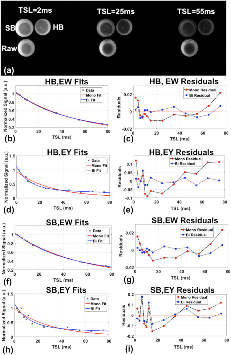
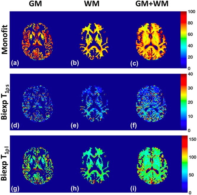
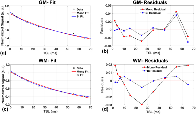
Detection of multiple relaxation pools using MRI is useful in a number of neuro-pathologies including multiple sclerosis (MS), Alzheimer's, and stroke. In this study we evaluate the feasibility of using T1ρ imaging for the detection of bi-exponential decays in the human brain. A prospective T1ρ imaging study was performed on model relaxation phantoms (eggs) and 7 healthy volunteers. The data was fitted using a single pool and a 2-pool model to estimate mono- and bi-exponential T1ρ maps, respectively. Bi-exponential decays were identified in the gray matter (GM) and white matter (WM) of the brain with 40.5% of GM, and 65.1% of WM pixels showing two T1ρ relaxation pools (significance level P < 0.05). Detection of T1ρ based bi-exponential decays in the brain provides complimentary information to T based contrast regarding the in vivo micro-environment in the brain.
使用 MRI 检测多个弛豫池在多种神经病理学中都很有用,包括多发性硬化症 (MS)、阿尔茨海默病和中风。在这项研究中,我们评估了使用 T1ρ 成像检测人脑双指数衰减的可行性。对模型弛豫体模(鸡蛋)和 7 名健康志愿者进行了前瞻性 T1ρ 成像研究。使用单池和双池模型对数据进行拟合,分别估计单指数和双指数 T1ρ 图谱。在大脑的灰质 (GM) 和白质 (WM) 中可以识别出双指数衰减,GM 中有 40.5%,WM 中有 65.1%的像素显示出两个 T1ρ 弛豫池(显著性水平 P<0.05)。在大脑中检测基于 T1ρ 的双指数衰减提供了关于大脑内体环境的与 T 相关对比的补充信息。